Treatment of Dural Solitary Fibrous Tumor/Hemangiopericytoma of Spine with a New Tyrosine Kinase Inhibitor Anlotinib Hydrochloride: A Case Report
- Corresponding Author:
- Wen-Xin Li
Departments of Oncology
Inner Mongolia People’s Hospital
Hohhot, Inner Mongolia, China
E-mail: wenxinli@mail.com
Abstract
The treatment options for high-grade dual Hemangiopericytoma (HPC) of the Central Nervour System (CNS) include surgical resection, radiation, and chemotherapy. Target therapy has no been reported in the literature. The choice of pharmaceutical target treatment for the Central Nervous (CNS) hemangiopericytoma is very limited. The pathogenesis of hemangiopericytoma is thought to relate to tumor angiogenesis and high expression of Vascular Endothelial Growth Factor (VEGF) and Platelet-Derived Growth Factor (PDGF) of tumor cells. The Tyrosine Kinase Inhibitor (TKI) that is proven to have potent inhibitory effects on VEGFR, PDGFR, FGFR, and c-Kit at the same time would be the ideal choice of the targeted therapy for hemangiopericytoma. In this article, we report a 29-year-old man with advanced high-grade hemangiopericytoma arising from the dura of the spinal cord received targeted therapy of this newly approved tyrosinase inhibitor Anlotinib Hydrochloride, which has the capability of multiple target points. Two weeks after the treatment, the size of the tumor was reduced significantly based on image analysis, and the patient’s compressive symptoms were improved considerably. This is the first case of high-grade hemangiopericytoma successfully managed with these newly approve tyrosine kinase inhibitors, a large scale clinical research on the targeted therapy of this tumor would be necessary to further understanding the pharmaceutical mechanisms of Anlotinib Hydrochloride on the tumor suppression of CNS hemangiopericytoma.
Keywords
Hemangiopericytoma; Tyrosinase inhibitor; Spinal cord; Anlotinib Hydrochloride.
Abbreviations
BCL 2: B-Cell Lymphoma 2; CNS: Central Nevous System; FGF: Fibroblast Growth Factor; NAB2: Nuclear Polyadenylated RNA-Binding Protein 2; WHO: World Health Organization; STAT: Signal Transducer and Activator of Transcription; PET-CT: Positron Emission Tomography-Computed Tomography; VEGF: Vascular Endothelial Growth Factor; PDGF: Platelet-Derived Growth Factor; SFT/HPC: Solitary Fibrous Tumor/Hemangiopericytoma
Introduction
Dural hemangiopericytoma is a rare tumor with metastatic capability. The low-grade tumor has a low rate of recurrence and better surgical treatment outcomes. In contrast, the high-grade tumors have aggressive growth ability, a high local recurrence rate, and distant metastasis. Local surgical control is less effective, radiotherapy is controversial, and available chemotherapy was ineffective [1,2]. The new treatment should be identified. According to the characteristics of tumor origin cells and the therapeutic targets covered by corresponding drugs, we explored the use of drugs and obtained better results. The tight connection between pericytes and vascular endothelial cells can affect angiogenesis, blood-brain barrier permeability, vascular system stability, immune regulation, cell migration, and differentiation [3].
Anlotinib hydrochloride has a strong inhibitory effect on VEGFR2, VEGFR3, PDGFR, and FGFR pathways. As we know that inhibiting VEGFR, PDGFR, and FGFR signaling pathways at the same time are less likely to cause bypass activation and restart angiogenesis than simply inhibiting one of them [4]. Anlotinib is the new tyrosinase inhibitor, with dual effects on tumor growth suppression and anagenesis inhibition. Anlotinib has been used in the treatment of advanced carcinoma of lung, kidney, and thyroid, as well as soft tissue sarcomas; the results are promising [5-7]. The multipart target point this agent is the favored pharmaceutical choice in the intervention in CNS HPC.
This is a case report of a new tyrosine kinase inhibitor that has dramatically reduced the tumor mass and improved the patient’s symptoms. This is the first successful targeted treatment for this tumor.
Case Presentation
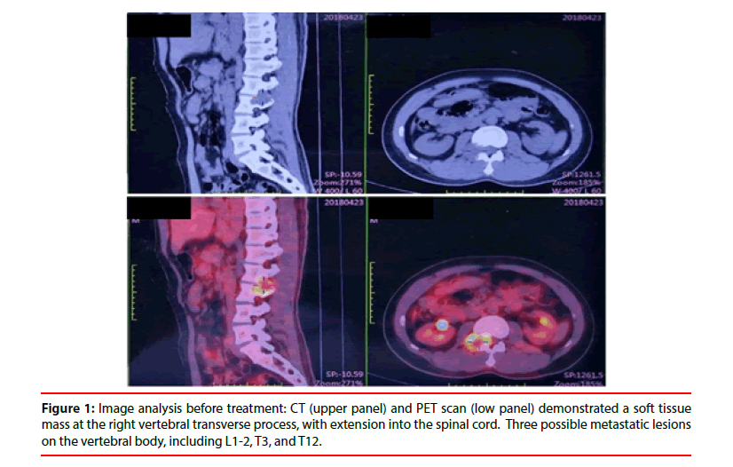
The patient is a 29-year-old Chinese male who presented to an outside hospital in April 2018 with complaints of worsening back pain of a two-month duration. He also complained of numbness and fatigue on the right leg. Completed PET-CT was done to reveal a soft tissue mass on the left thoracic cavity with uneven metabolism. The mass lesion has no clear borders with the precardiac sac, left diaphragm, and left pleura. The findings were suggestive of a malignant neoplasm. A second soft tissue mass was found at the right vertebral transverse process, which showed extension into the spinal cord. There were possible metastatic lesions noted on the vertebral body, including L1-2, T3, and T12 (Figure 1).
Biopsy of the peri-vertebral body soft tissue was done, and the pathology examination demonstrated a small round cell malignant neoplasm associated with extensive necrosis. Immunohistochemistry studies excluded the possibility of lymphoproliferative disorders, epithelial neoplasm, rhabdomyosarcoma, and neuroendocrine tumor; however, Ewing sarcoma could not be excluded.
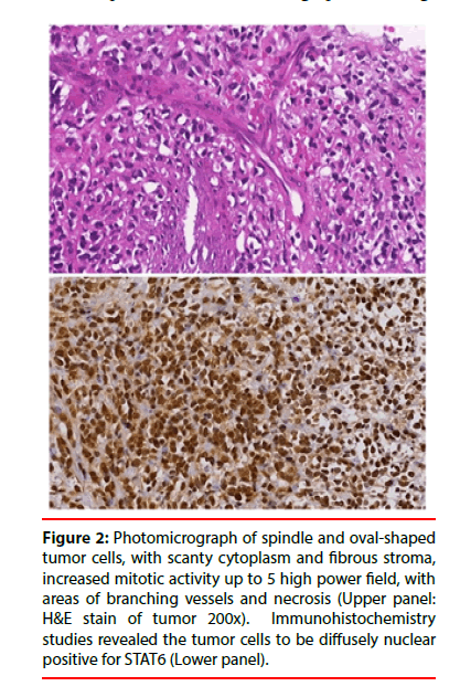
The patient came to our hospital for further evaluation. On admission, physical examination revealed severe pressure pain on the spinous process at L1 to L3, numbness of the right lower limb with a normal deep sensation of bilateral lower limbs. The muscle strength was approximately 3/5 for the bilateral quadriceps; accelerated knee reflex and tendon reflex was noted on both sides. Bilateral Hoffmann sign was negative, bilateral Babinski sign was positive, and bilateral ankle spasm was positive. The patient preferred conservative radiation therapy and sodium bi-phosphates over surgical intervention to relieve the compressive symptom. Meanwhile, repeat biopsies on the soft tissue mass were performed on both the peri vertebral body and the thoracic cavity. The pathology examination of the mass from the peri vertebral body demonstrated a mesenchymal neoplasm with increased cellularity. The cells were arranged in irregular fascicles with focal branching vessels that were seen with perivascular hyalinization. The tumor is seen intermingled with bone fragments, and focal necrosis is noted. At high power, the tumor cells appeared to be spindle and oval-shaped, with scanty cytoplasm and fibrous stroma, increased mitotic activity up to 5 high power field, with areas of necrosis. Immunohistochemistry studies revealed the tumor cells to be diffusely nuclear positive for STAT6 (Figure 2).
Figure 2: Photomicrograph of spindle and oval-shaped tumor cells, with scanty cytoplasm and fibrous stroma, increased mitotic activity up to 5 high power field, with areas of branching vessels and necrosis (Upper panel: H&E stain of tumor 200x). Immunohistochemistry studies revealed the tumor cells to be diffusely nuclear positive for STAT6 (Lower panel).
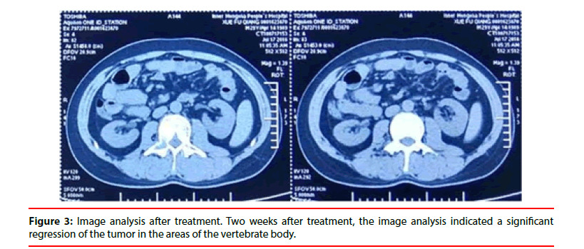
Based on the morphologic features and immunophenotypic profile (the tumor cells are BCL2 positive, Ki 67 more than 40%), the tumor is consistent with CNS solitary fibrous tumor hemangiopericytoma, grade 3. The transthoracic CT-guided biopsy from the thoracic cavity revealed only fibrocartilage and might not represent the mass lesion. The clinical presentation and radiographic findings were supportive of the diagnosis of peri spinal dura hemangiopericytoma with metastasis to bone. Upon discussing the results with the patient, the decision was made to treat the tumor with Anlotinib Hydrochloride, 12 mg daily. Two weeks after treatment, the image analysis indicated a significant regression of the tumor in the areas of the vertebrate body along with significantly improved clinical symptoms (Figure 3).
The patient tolerated the treatment very well, except for mild stomatitis. No significant alterations of the peripheral blood parameters, blood pressure, urine analysis, and the functions of the liver and kidney are noted. Following up at six months after treatment, the patient has no neural compressing symptoms and no radiographic evidence of tumor recurrence.
Discusion and Conclusion
Solitary Fibrous Tumor (SFT) and hemangiopericytoma in the central nervous system show similar histologic and immune phenotypic features as well as overlapping ultra-structure changes [8,9]. The most important recent finding is that both tumors have fusion gene NAB2-STAT6. The World Health Organization (WHO) classified CNS hemangiopericytoma as Solitary Fibrous Tumor/ Hemangiopericytoma (SFT/HPC). The tumor is rare and is related to mesenchymal fibroblast cells, and is marginally malignant with the ability for local invasion and metastasis [10]. Most central nervous system HPC originated from the dura, and it is extremely rare in the spinal cord. The outside hospital PET-CT studies of this patient suggested the lesion protrude into the spinal cord from the peri-vertebral area; however, it appeared to be more reasonable than the tumor was originally from the dura of the spinal cord and invaded to the peri vertebral body and metastasized to distant bone.
Signal Transducer and Activator of Transcription (STAT) transcription factors are a family of endocytoplasm proteins with similar structures that participate in gene transcription regulation when binding to nuclear DNA once the proteins are activated [11]. It plays an important role in various physiological and pathologic processes, including cell immune reactions, cell proliferation and differentiation, and tumorigenesis. STAT6 is one of the family members of this protein. The fusion gene of NAB2-STAT6 and its product is a specific molecular marker for SFT/HPC and a tumor oncogene to develop solitary fibrous tumor/hemangiopericytoma [12]. The tumor suppressor gene NAB2 acquired an activation zone on STAT6 via gene fusion. In this process, the ERG signal cycle is persistently active and eventually result in cell proliferation and tumor formation. Besides, the fusion gene of NAB2- STAT6 may serve as a new targeted point in tumor therapy [13].
During embryonic development, PDGF-β released by endothelial cells binds to PDGFR-β on pericytes, promoting angiogenesis and cell development. When PDGF-β is blocked, the activity of pericytes will be decreased, and the number of migration around the endothelium will be reduced, which will affect angiogenesis. It is well known that VEGFR, PDGFR, and FGFR signal pathways are the primary mechanism of tumor angiogenesis. At the same time, the signaling pathways of angiogenesis can achieve crosstalk through the regulation of ERK, Akt, and PI3K in the downstream signal transmission process, which means that inhibiting VEGFR, PDGFR, and FGFR signaling pathways at the same time are less likely to cause bypass activation and restart angiogenesis than simply inhibiting one of them [4]. VEGF/PDGF/FGF is a major tumor angiogenesis signal pathway. PDGF plays a vital role in the modulation of pericytes and related vascular proliferation. There is cross talk in all three-signal pathways; therefore, there is a limited effect on tumor angiogenesis when only one or two pathways are blocked, and the effects, if any, are short-lived. Anlotinib Hydrochloride is a newly developed kinase inhibitor, competing for ATP binding point on VEGFR/PDGFR/FGFR/C-KIT, and block the subtract phosphorylation and signal transduction simultaneously, and persistently suppress all four-tyrosine kinase phosphorylation in the process of tumor proliferation [6,7]. Besides, this new treatment agent has focused on the target point, lower IC 50, and lower side effects as compared to other common tyrosine kinase inhibitors.
Regarding the giant mass in the thoracic cavity, no significant changes were revealed by CT studies after treatment (Fig not shown). This tumor is most likely a synchronous solitary fibrous tumor, and similar cases have also been reported previously [5,6]. In general, solitary fibrous tumor in the thoracic cavity is solid, with pseudo capsule and firm consistency, and may be associated with hemorrhage, necrosis, and calcifications. It is possible that the dura/CNS high-grade hemangiopericytoma with potent tumor angiogenesis activity and, therefore, responds to the TKI intervention effectively, while the tumor in the thoracic cavity, likely an indolent solitary fibrous tumor, responds less to the treatment.
Declarations
Ethics approval to use Aniotinib hydrochloride was granted by the ethics committee of clinical investigation of Inner Mongolia Autonomous region People’s Hospital. Consent for publication are done.
Availability of Data and Materials
The data sets used and/or analyzed during the current study are available from the corresponding author on reasonable request.
Authors’ Contributions
Wei Luan and Lan Yu patient information collecting, data analysis and manuscript drafting, etc. final reading of the manuscript. Zheng Liu pathology examination and final reading of the manuscript. Wen-Xin Li case treatment design and final reading of the manuscript.
Competing Interests
The authors declare that they have no competing interests in this section.
Funding
No funding is available for this case report.
References
- Liu H, Yang A, Chen N. Hemangiopericytomas in the spine: Clinical features, classification, treatment, and long-term follow-up in 26 patients. Neurosurgery 72, 16-24 (2013).
- Ecker R, Marsh W, Pollock B. Hemangiopericytoma in the central nervous system: Treatment, pathological features, and long-term follow up in 38 patients. J Neurosurg 98, 1182-1187 (2003).
- Bergers G, Song S. The role of pericytes in blood-vessel formation and maintenance. Neuro Oncol 7, 452-464 (2005).
- Song M, Finley SD. ERK and Akt exhibit distinct signaling responses following stimulation by pro-angiogenic factors. Cell Commun Signal 18, 114 (2020).
- Si X, Zhang L, Wang H et al. Management of anlotinib-related adverse events in patients with advanced non-small cell lung cancer: Experiences in ALTER-0303. J Thoracic cancer 10, 551-556 (2019).
- Wu G, Cheng Y, Shi Y, et al. Anlotinib as a third-line therapy in patients with refractory advanced non-small-cell lung cancer: A multicentre, randomised phase II trial (ALTER0302). Br J Cancer 118, 654-661 (2018).
- Song Z, Yu X, Lou G, et al. Salvage treatment with anlotinib for advanced non-small-cell lung cancer. Onco Targets Ther 10, 1821-1825 (2017).
- Savary C, Rousselet M, Michalak S. Solitary fibrous tumors and hemangiopericytomas of the meninges: Immunophenotype and histoprognosis in a series of 17 cases. Ann Pathol 36, 258-267 (2016).
- Dan R, Wu Y, Kalyanasundaram S. Identification of recurrent NAB2-STAT6 gene fusions in solitary fibrous tumor by integrative sequencing J Nat Genet 45, 180-185 (2013).
- Dan RR, Yi-Mi W, Shanker KS, et al. Identification of Recurrent NAB2-STAT6 Gene Fusions in Solitary Fibrous Tumor by Integrative Sequencing. Nat Genet 45, 180-185 (2013).
- Wang K, Zhang S, Shi L. The 2016 World Health Organization classification of tumors of the central nervous system: A summary. J Acta Neuropathol 131, 803-820 (2016).
- Yoshimoto M, Kurihara H, Fujii H. Theragnostic imaging using radiolabeled antibodies and tyrosine kinase inhibitors. https://pubmed.ncbi.nlm.nih.gov/25874259/
- Petrella F, Monfardini L, Musi G, et al. Synchronous pleuro-renal solitary fibrous tumors: A new clinical-pathological finding. Minerva Chir 64, 669-671 (2009).


