Relationship of Morphological Changes of the First Molar Pulp Chamber and Mineralization of Developing Third Molar with Cervical Vertebral Maturation on Panoramic Radiographs and Lateral Cephalograms
- Corresponding Author:
- Maruf Noruzi
Department of Oral and Maxillofacial Surgery, Dental Faculty, Urmia University of Medical Science, Urmia, Iran
E-mail: maruf.noruzi@yahoo.com
Abstract
ABSTRACT
Objectives: The study aimed to assess the relationship of morphological changes of the first molar pulp chamber and mineralization of developing the third molar with Cervical Vertebral Maturation (CVM) on panoramic radiographs and lateral cephalograms.
Materials and Methods: This descriptive cross-sectional study evaluated 410 panoramic radiographs and lateral cephalograms of patients between 8-18 years in Urmia city, Iran. The CVM was evaluated using the Hassel and Farman method while third molar mineralization was evaluated using the Demirjian’s Index (DI). The morphology of the mandibular left first molar pulp chamber was evaluated using the Tooth Crown Index (TCI). Spearman’s correlation coefficient was calculated to assess the correlation between the variables. Comparisons were made using the chi-square test and one-way ANOVA.
Results: The chronological age of males and females showed highly significant correlations with DI and CVM index, respectively (0.874, 0.810, 0.882 and 0.800). A high correlation existed between DI and CVM index, which was higher in males (0.901) than females (0.790). No significant correlation was noted between TCI and chronological age or CVM index in males or females (p>0.05).
Conclusion: The third molar mineralization stage determined on panoramic radiographs and lateral cephalograms can be clinically used to assess skeletal maturity and estimate the chronological age.
Keywords
Cephalometry; Cervical vertebra; Dental mineralization; Panoramic radiography
Introduction
Age estimation plays an important role in forensic medicine, pediatric endocrinology, and clinical dentistry particularly orthodontics [1,2]. Age estimation in children and adolescents is often performed by the use of chronological, skeletal, dental, anthropometric or physiologic methods [3]. Chronological age is the most simple and most commonly used measure to assess an individual’s growth status. However, considerable variability in the growth and development of individuals with the same chronological age renders it an unreliable index for assessment of growth potential. This led to the use of physiological age, instead of chronological age, which makes the prediction of growth potential more specific [4].
Assessment of developmental skeletal changes is among the most commonly used methods for estimation of growth potential, which is more accurate and clinically more efficient than using the chronological age for this purpose [4]. During growth and development, bones undergo a series of changes. The order of the of a specific bone is relatively the same in different individuals; however, the timing of these changes may vary among them, depending on their biological clock [3]. Hand wrist radiography is among the most commonly used methods for assessment of skeletal growth and development. In radiographic age estimation, bones exhibit less variability compared to other indices. Moreover, some concerns exist regarding the unnecessary radiation exposure in hand wrist radiography [4]. Thus, assessment of skeletal maturity by evaluation of the cervical vertebrae has recently gained increasing popularity because the cervical vertebrae are conventionally visualized on lateral cephalograms normally requested for orthodontic treatment and therefore, the patients are not subjected to unnecessary radiation exposure. Evidence shows that the assessment of the developmental stage of the cervical vertebrae can be a reliable technique for the determination of skeletal age [4].
Tooth structure is a unique parameter for the estimation of dental age. The development of dental tissues is less affected by endocrine disorders and nutritional variations than other tissues [1]. Two main approaches in the determination of dental age include the age of tooth eruption and pattern of dental calcification. The timing of tooth eruption used to be a popular index for age estimation but it is no longer considered accurate [3]. In contrast, the pattern of calcification and tooth formation is a long process with several phases, each having its own definition and characteristics [3].
Panoramic radiographs are also available as routine dental radiographs for many patients and therefore, may be used for assessment of dental mineralization and its correlation with skeletal age [5]. A strong correlation exists between the stage of dental mineralization and skeletal age [6]. Evidence shows that the first premolar and second molar teeth in females and the first premolar, canine and second molar teeth in males are the best growth predictors. The second molars are the most accurate for this purpose in both males and females [6]. The level of mineralization of the third molar root can also be used for age estimation [7]. After the eruption of permanent second molars at the age of 12 to 13 years, age estimation becomes more complicated. However, third molars are forming in this age range and may be used for age estimation. Third molars erupt into the oral cavity at the age of 17 to 21 years. They have distinct phases of development determined based on their root formation, which starts at the age of 14 years [3]. Evidence shows a positive correlation between the development of third molars and the overall growth pattern of individuals [7].
Over time, the size of the pulp chamber diminishes due to the formation of secondary dentin. Clinical changes in the size of the pulp chamber are due to pulpal recession, which is detectable on radiographs. The size of the pulp chamber, which is expected to decrease over time, has a significant correlation with chronological age. By aging, the size of the pulp chamber significantly decreases and calcification of the coronal and radicular pulp occurs. Evidence shows that the reduction in the size of the pulp chamber can serve as an important index for age estimation [1].
Given that a significant correlation exists between the developmental stage of third molars and overall growth, third molars may be used for growth prediction. On the other hand, given that a correlation exists between the morphological changes of the pulp chamber and skeletal maturity, it can be used for treatment planning at this age range. This study, therefore, aimed to assess the relationship of morphological changes of the mandibular first molar pulp chamber and mineralization of developing the third molar with Cervical Vertebral Maturation (CVM) on panoramic radiographs and lateral Cephalograms.
Materials and Methods
This descriptive cross-sectional study evaluated patients between 8 to 18 years who had both panoramic radiographs and lateral Cephalograms as a prerequisite for their orthodontic treatment. Patients were selected using convenience sampling. In this study, five variables namely age, gender, mineralization of the third molar, Tooth Crown Index (TCI), CVM and the correlation among them were evaluated.
All panoramic and lateral cephalometric radiographs had been taken by one oral and maxillofacial radiologist using the Planmeca system (Scara 3; Planmeca, Helsinki, Finland). The head position and exposure settings were adjusted individually for each patient.
The inclusion criteria were the availability of panoramic radiograph and lateral Cephalograms of patients with optimal quality, absence of systemic diseases and local conditions affecting tooth development, no root resorption or calcification, no dental anomaly and knowledge about chronological age and sex of patients.
The exclusion criteria were a distortion of panoramic images or lateral Cephalograms, hypodontia or extensive pathological lesions affecting the mandibular left quadrant, presence of caries or restorations or endodontic treatment of the first molar tooth, visualization of thyroid collar on lateral Cephalograms and presence of buccal or lingual dental tilting.
A total of 410 panoramic and lateral cephalometric radiographs met the eligibility criteria and were visually evaluated by two independent observers. Inter-examiner calibration was performed to standardize the interpretation of images by the two observers by evaluating 10% of the radiographs.
Age (year and month) and sex of patients were first retrieved from patient files and recorded.
▪ Assessment of third molar mineralization
The developmental stage of mandibular left third molar was determined using the Demirjian’s Index (DI), which classified tooth development into 8 stages from A to H as follows [2] (Figure 1):

Figure 1: Mineralization stages of mandibular left third molar according to the Demirjian’s index (A): initiation of calcification; (B): cusp formation; (C): initiation of dentin deposition; (D): initiation of root formation; (E): initiation of root bifurcation; (F): funnel-shaped roots; (G): open apex and; (H): apex closure.
• In single-rooted and multi-rooted teeth, calcification starts at the superior border of the crypt in the form of one or more inverted cones with no attachment between them (Figure 1, stage A)
• By connecting the calcified regions, one or more cusps form and determine the morphology of the tooth crown (Figure 1, stage B)
• Half of the crown is completed and the pulp chamber forms. Dentin deposition starts (Figure 1, stage C)
• Tooth crown has formed to the level of the Cementoenamel Junction (CEJ) and the pulp chamber has a trapezoidal shape. Root starts to form (Figure 1, stage D)
• Root bifurcation starts to form; however, the root is still shorter than the crown (Figure 1, stage E)
• The root end is funnel-shaped and the root length is equal or longer than the crown length (Figure 1, stage F)
• Root canal walls are parallel and the apex is open (Figure 1, stage G)
• The apex is completely closed and the periodontal ligament has an equal width around the root and apex (Figure 1, stage H)
▪ Assessment of the pulp chamber morphology
For assessment of the pulp chamber morphology of the mandibular left first molar, a line was drawn from the CEJ to hypothetically divide the anatomical crown and root in half. Crown Height (CH) was then vertically measured from the CEJ to the tip of the longest cusp. The coronal Pulp Cavity Height (CPCH) was vertically measured from the cervical line to the tip of the highest pulpal horn.
The Tooth Crown Index (TCI) was calculated using the formula 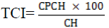
The TCI was separately calculated for each patient (Figure 2).

Figure 2: The Tooth Crown Index (TCI) calculation.
▪ Assessment of CVM index
The cervical vertebrae were evaluated (I) based on the concavity of the inferior border of C2, C3, C4, and (II) shape of C3 and C4.
For assessment of the inferior border of C2, C3 and C4, the anterior-inferior and posteriorinferior border of each vertebra were connected by a straight line. Another line was drawn a tangent to the approximate center of each vertebra (by forming an angle) and the distance from the straight line to the line drawn at the center determined the concavity of the inferior border of the cervical vertebra. If the concavity depth at the inferior border was >1 mm, the border was considered concave; otherwise, it was considered smooth or flat (Figures 3 and 4).

Figure 3: Evaluation the inferior border of cervical vertebrae concavity.

Figure 4: Geometrical shape of cervical vertebrae: Flat inferior border and concave inferior border.
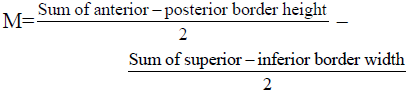
Assessment of the geometrical shape of C3 and C4 was done using the formula proposed by McNamara et al. [8] by measuring the height of the anterior and posterior border and the superior and inferior width of each vertebra (Figure 5). The M value in the following formula determines the geometrical shape of each vertebra.

Figure 5: Assessment of the geometrical shape of C3 and C4.
After determining the geometrical shape and concavity/smoothness of the inferior border of each vertebra, the CVM stage was determined using the following classification (Figure 6):

Figure 6: The CVM stage.
CS1: The inferior border of all three vertebrae is smooth and the body of C3 and C4 is trapezoidalshaped. The mandibular growth spurt occurs 2 years after this stage.
CS2: The inferior border of C2 is concave and the body of C3 and C4 is trapezoidal-shaped; the mandibular growth spurt occurs 1 year after this stage.
CS3: The inferior border of C3 is concave and the body of C3 and C4 has a trapezoidal or horizontal rectangular shape; this stage is simultaneous with the mandibular growth spurt.
CS4: The inferior border of C2, C3, and C4 are concave and the body of C3 and C4 has a horizontal rectangular shape. The mandibular growth spurt has passed by 1-2 years.
CS5: The inferior border of C2, C3, and C4 are concave and the body of C3 or C4 is squareshaped. The mandibular growth spurt has ended one year earlier.
CS6: The inferior border of C2, C3, and C4 are concave and the body of at least C3 or C4 has a vertical rectangular shape. The mandibular growth spurt has ended 1 to 2 years earlier.
The lateral geometrical shape of the cervical vertebrae (Figure 7) was determined using Romexis software (Planmeca, Helsinki, Finland). Data regarding DI, CVM and TCI were analyzed using Excel software and by use of descriptive and inferential statistics (Figure 8). The Kolmogorov-Smirnov test was used to assess the normality of data distribution. All data had non-normal distribution (p<0.05) except for the TCI (p=0.119). Spearman’s correlation coefficient was used to assess the relationship between non-parametric variables. One-way ANOVA was used to analyze TCI. Inter-observer reliability was evaluated by calculating the intra-class correlation coefficient.

Figure 7: Assessment of the shape of the vertebral bodies. (a): square-shaped (-1<M<+1); (b): vertical rectangular shape (M>+1); (c): horizontal rectangular or trapezoidal shape (M<-1).

Figure 8: Determining the lateral geometrical shape of the cervical vertebra (by coding in Excel software).
Results
The inter-observer reliability was found to be excellent. A total of 410 patients between 8-18 years were evaluated; out of which, 131 were males and 279 were females. Table 1 shows the frequency of male and female patients in different age groups.
| Age group (yrs.) | Males (n) | Females (n) |
|---|---|---|
| 8-9 | 15 | 16 |
| 9-10 | 13 | 22 |
| 10-11 | 14 | 26 |
| 11-12 | 23 | 58 |
| 12-13 | 12 | 44 |
| 13-14 | 24 | 32 |
| 14-15 | 12 | 33 |
| 15-16 | 5 | 16 |
| 16-17 | 7 | 16 |
| 17-18 | 6 | 16 |
Table 1: Frequency of male and female patients in different age groups.
Table 2 shows the mandibular third molar mineralization stage (DI) according to the chronological age and gender of patients. Assessment of the correlation of the third molar mineralization stage and chronological age with the chi-square test revealed a significant association between them (p<0.05). Table 3 shows the mandibular third molar mineralization stage based on gender. The Spearman’s correlation coefficient showed a high correlation between the third molar mineralization stage and chronological age in both males and females (p=0.00, correlation coefficient: 0.810 in females and p=0.00, correlation coefficient: 0.874 in males).
| Age | 8-9 | 9-10 | 10-11 | 11-12 | 12-13 | 13-14 | 14-15 | 15-16 | 16-17 | 17-18 | ||||||||||
|---|---|---|---|---|---|---|---|---|---|---|---|---|---|---|---|---|---|---|---|---|
| Sex/DI | F | M | F | M | F | M | F | M | F | M | F | M | F | M | F | M | F | M | F | M |
| A | 14 | 10 | 8 | 6 | 3 | 5 | 8 | 1 | 0 | 0 | 0 | 0 | 0 | 0 | 0 | 0 | 0 | 0 | 0 | 0 |
| B | 2 | 5 | 12 | 7 | 15 | 8 | 17 | 7 | 6 | 0 | 4 | 0 | 0 | 0 | 0 | 0 | 0 | 0 | 0 | 0 |
| C | 0 | 0 | 2 | 0 | 8 | 1 | 21 | 12 | 21 | 9 | 10 | 11 | 9 | 3 | 0 | 0 | 0 | 0 | 0 | 0 |
| D | 0 | 0 | 0 | 0 | 0 | 0 | 9 | 1 | 7 | 1 | 6 | 3 | 11 | 3 | 7 | 0 | 1 | 1 | 0 | 0 |
| E | 0 | 0 | 0 | 0 | 0 | 0 | 3 | 2 | 8 | 2 | 10 | 5 | 8 | 3 | 5 | 3 | 3 | 2 | 3 | 0 |
| F | 0 | 0 | 0 | 0 | 0 | 0 | 0 | 0 | 1 | 0 | 1 | 1 | 4 | 3 | 1 | 1 | 3 | 1 | 1 | 3 |
| G | 0 | 0 | 0 | 0 | 0 | 0 | 0 | 0 | 1 | 0 | 1 | 4 | 1 | 0 | 2 | 1 | 8 | 3 | 7 | 3 |
| H | 0 | 0 | 0 | 0 | 0 | 0 | 0 | 0 | 0 | 0 | 0 | 0 | 0 | 0 | 1 | 0 | 1 | 0 | 5 | 0 |
Table 2: Frequency of mandibular third molar mineralization stage (DI) according to the chronological age and gender of patients.
| DI/Sex | Male | Female | ||
|---|---|---|---|---|
| Mean | Std. deviation | Mean | Std. deviation | |
| A | 10.24 | 1.217 | 9.94 | 1.478 |
| B | 12.56 | 1.219 | 11.43 | 1.291 |
| C | 13.08 | 1.052 | 12.79 | 1.275 |
| D | 14.33 | 1.414 | 14.67 | 1.506 |
| E | 15.53 | 1.546 | 15.62 | 1.659 |
| F | 16.22 | 1.563 | 16.64 | 1.502 |
| G | 16.09 | 1.758 | 16.80 | 1.399 |
| H | - | - | 17.57 | 0.787 |
Table 3: Mean age of mineralization of mandibular third molar based on gender.
Table 4 presents the frequency of CVM index according to the chronological age and gender of patients. The chi-square test showed a significant correlation between the third molar mineralization stage and chronological age (p<0.05). Table 5 shows the mean CVM index in males and females. According to the Spearman’s correlation coefficient, a high correlation existed between CVM index and chronological age in both males (p=0.00, correlation coefficient: 0.882) and females (p=0.00, correlation coefficient: 0.800).
| Age | 8-9 | 9-10 | 10-11 | 11-12 | 12-13 | 13-14 | 14-15 | 15-16 | 16-17 | 17-18 | ||||||||||
|---|---|---|---|---|---|---|---|---|---|---|---|---|---|---|---|---|---|---|---|---|
| Sex/CVM | F | M | F | M | F | M | F | M | F | M | F | M | F | M | F | M | F | M | F | M |
| CS1 | 13 | 15 | 7 | 13 | 11 | 4 | 8 | 2 | 0 | 0 | 0 | 0 | 0 | 0 | 0 | 0 | 0 | 0 | 0 | 0 |
| CS2 | 3 | 0 | 13 | 0 | 9 | 8 | 15 | 10 | 2 | 1 | 0 | 3 | 0 | 0 | 0 | 0 | 0 | 0 | 0 | 0 |
| CS3 | 0 | 0 | 2 | 0 | 2 | 0 | 10 | 8 | 5 | 8 | 4 | 2 | 1 | 2 | 0 | 0 | 0 | 0 | 0 | 0 |
| CS4 | 0 | 0 | 0 | 0 | 4 | 2 | 20 | 2 | 20 | 1 | 7 | 12 | 8 | 2 | 3 | 0 | 2 | 2 | 2 | 0 |
| CS5 | 0 | 0 | 0 | 0 | 0 | 0 | 5 | 1 | 15 | 2 | 20 | 7 | 19 | 7 | 11 | 3 | 13 | 4 | 9 | 4 |
| CS6 | 0 | 0 | 0 | 0 | 0 | 0 | 0 | 0 | 2 | 0 | 1 | 0 | 5 | 1 | 2 | 2 | 1 | 1 | 5 | 2 |
Table 4: Frequency of CVM index and its correlation with chronological age and gender.
| DI/Sex | Male | Female | ||
|---|---|---|---|---|
| Mean | Std. deviation | Mean | Std. deviation | |
| CS1 | 10.59 | 1.131 | 10.15 | 1.443 |
| CS2 | 11.95 | 0.999 | 11.00 | 1.082 |
| CS3 | 12.90 | 0.968 | 12.42 | 1.248 |
| CS4 | 13.86 | 1.526 | 13.33 | 1.649 |
| CS5 | 15.32 | 1.679 | 14.99 | 1.719 |
| CS6 | 16.67 | 0.211 | 15.88 | 1.784 |
Table 5: Mean CVM index in males and females.
Assessment of the correlation of third molar mineralization and CVM revealed that DI B in both males and females had the highest frequency in CVM2, which was before the growth spurt. The DI B in females and DI C in males had a significant correlation with CVM1, which was simultaneous with the growth spurt. The chisquare test revealed a significant correlation between the stage of third molar mineralization and chronological age (p=0.00). The correlation coefficient in males (0.901) was higher than that in females (0.790).
One-way ANOVA showed no significant correlation between the mean changes in the size of the pulp chamber in mandibular first molar (TCI) and chronological age (p=0.893 in females and p=0.738 in males). No significant correlation was noted between TCI and CVM index in males (p=0.931) or females (p=0.155) either.
Discussion
The stage of mineralization and development of teeth is widely used for growth prediction and determination of growth spurt, which is highly important in orofacial orthopedics [4]. The advantage of radiographic assessment of dental maturation is that it can be easily performed during the course of routine dental treatments [7].
In general, two methods are used to determine the chronological age based on dentition: (I) age estimation according to the time of tooth eruption and (II) use of tooth mineralization pattern [3]. The disadvantage of age estimation methods based on tooth eruption is that the time of eruption cannot be accurately determined and is affected by environmental factors such as systemic diseases and nutrition; therefore, this method is not highly reliable [7].
Several methods have been proposed for the estimation of chronological age according to the pattern of tooth mineralization [7]. Demirjian et al. [9] introduced an index to determine the dental maturation stage according to the dental mineralization pattern. DI is simple and provides an accurate description of the mineralization process for each stage, which leads to the highest intra-observer reliability [9]. The accuracy and reliability of the DI have been previously confirmed in the Iranian population [10]. The modified DI focuses on the root of the third molar tooth. In this index, proposed by Orhan et al., [11] the allocated score for cases without a mandibular third molar or presence of tooth bud radiolucency is zero.
In the present study, the mandibular left third molar was evaluated for the assessment of dental mineralization. It should be noted that the standard DI is for the permanent mandibular left teeth [4]. Thus, the mandibular left third molar was evaluated in our study.
Root formation and apex closure in all permanent teeth, except for third molars, often accomplished by the age of 14 years while Bolanos et al. [12] showed that closure of the mandibular third molar root apex occurs at the age of 18 years. Thus, using the third molar mineralization stage as an index is unique since it occurs over a long period of time and continues to early adulthood; therefore, it is less commonly affected by environmental factors due to its late eruption. Third molar development has a wide variation in different populations and can be reliable for age estimation in different races using the DI [7]. Our findings revealed a significant correlation between the mineralization stage of the third molar according to the DI and chronological age. These results were in line with those of Araujo et al., [13] Lewis et al., [14] and Orhan et al., [11] who showed a significant correlation between third molar mineralization stage and chronological age. The difference in the results of studies is attributed to the time of completion of third molar mineralization. Some studies have reported earlier completion of third molar mineralization in males while some others reported earlier completion of third molar mineralization in females [15]. Araujo et al., [13] stated that the absolute velocity of mineralization was the same in males and females and small differences in the dental development of females are attributed to the smaller size of teeth. No significant difference has been reported in the third molar mineralization stage in the right and left sides, or the maxilla and mandible [16-18].
Aside from the method of age estimation, skeletal age has the highest relationship with orthodontic treatment and diagnosis. This relationship allows the determination of chronological age according to skeletal maturation [19]. Hassel and Farman [20] modified the CVM index introduced by Lamparski et al., [21] and used the body of C2, C3 and C4 on lateral cephalograms to determine the skeletal age. CVM index is the gold standard for the assessment of cervical vertebrae [22]. Considering the easy assessment of CVM on lateral cephalograms and the prevention of unnecessary radiation exposure, the use of handwrist radiography is being increasingly limited [7]. In the present study, we used the CVM on lateral cephalograms routinely requested by orthodontists for orthodontic treatment. Our results indicated a high agreement between the chronological age and skeletal maturation of cervical vertebrae in both males and females; this correlation was higher in males. This finding was in agreement with that of Abesi et al., [4] in an Iranian population and Baidas [23] in Saudi Arabia. Uysal et al., [24] in Turkey and Stiehl et al., [25] in Germany found a weak correlation between chronological age and CVM. The difference in the results of studies may be attributed to different races, geographical location, age group, sample size, and sample selection criteria [4].
Assessment of the correlation between CVM index and stage of third molar mineralization according to the DI revealed a high correlation between these two variables which was higher in males than females. Singh et al., [26] reported similar results in an Indian population while Felemban et al., [27] reported a moderate to low correlation in Saudi Arabia. This controversy can be due to racial differences [4]. Evidence shows that calcification of mandibular second molar and canine teeth has a strong correlation with skeletal maturation [7]. In our study, the DI B had the highest frequency in CVM2 in both males and females, which is simultaneous with the period right before the growth spurt. DI B in females and DI C in males had a correlation with CVM3, which is simultaneous with the growth spurt period. This result was in accordance with that of Fishman [28]. These findings indicate that the formation of third molars occurs sooner in females than males. The same results were reported by Sisman et al., [29] Golovcencu et al., [30] and Solari and Abramovitch [31].
Age estimation becomes complicated after the eruption of the third molars. Drusini et al. [32] used the TCI for age estimation. They used mandibular teeth for higher clarity on radiographs. Assessment of the correlation between the TCI of the first molar and chronological age and CVM in 8-18-year-olds revealed no significant correlation. Previous studies showed a significant correlation between the TCI and chronological age. Kvaal et al., [33] Drusini et al., [32] and Morse et al. [34] found a significant correlation between TCI and chronological age. Drusini et al., [32] reported a negative correlation coefficient between TCI and chronological age, indicating an inverse correlation between the two. In other words, TCI decreases by aging due to dentin deposition. The difference in the results of studies may be attributed to the different age ranges of patients. In young individuals, dentin deposition is minimal and therefore, small changes in the size of the pulp chamber do not reach statistical significance.
Considering the genetic and racial differences, the results of this study cannot be generalized to other populations. Similar future studies in other parts of Iran are required in order to be able to perform a meta-analysis and apply the results to routine dental practice in the Iranian population.
Conclusion
According to the obtained results, the mandibular third molar mineralization stage can be used for chronological age estimation in 14 to 18-year-old Iranians. Considering the need for panoramic and lateral cephalometric radiographs for routine orthodontic treatment, these radiographs can be used for age estimation and determination of skeletal maturity in orthodontic patients to prevent unnecessary radiation exposure.
References
- Haavikko K. Tooth formation age estimation on a few selected teeth a simple method for clinical use. Proc Finn Dent Soc 70,15-19 (1974).
- Imanimoghadam M, Bagherpour A, Tohidi E, et al. Age assessment of developmental stage of permanent mandibular teeth using the Demirjian method. J Mash Dent Sch 35(1), 9-16 (2011).
- Mirhaishemi AH, Jabbarian R. The use of dental radiography in age determination: Concepts and methods. J Dent Med 27(2), 137-143 (2014).
- Abesi F, Fattahi S, Haghanifar S, et al. The agreement of chronological age and cervical vertebrae morphologhy in lateral cephalogram in a selected iranian populatin. J Mash Dent Sch 39(1), 61-70 (2015).
- Jacome Lopes L, Oliveira Gamba T, Augusta Portella M, et al. Utility of panoramic radiography for identificatin of the pubertal growth period. Am J Orthod Dentofacial Orthop 149(4), 509-515 (2016).
- Nishit M, Patel D, Mehta F, et al. Evaluation of skeletal maturation using mandibular third molar development in Indian adolescents. J forensic Dent Sci 8(2), 112 (2016).
- Mustafaa S, Anekar J, Devang Divakar D, et al. Evaluation of dental and skeletal maturity using digital panoramic radiographs and digital cephalograms. Asian Biomedicine 9(3), 335-342 (2017).
- McNamara Jr JA, Franchi L. The cervical vertebral maturation method: A user's guide. The Angle Ortho 88(2), 133-143 (2018).
- Demirjian A, Goldstein H, Tanner JM. A new system of dental age assessment. Hum Biol 45(2), 211-227 (1973).
- Sheykhi M, Ghodoosi A, Ghadipasha M, et al. Radiographic survey of third molars development in relation to chronological age in iranian population. J Kerman Univ Med Sci 14(3), 195-202 (2007).
- Orhan K, Ozer L, Orhan AI, et al. Radiographic evaluation of third molar development in relation to chronological age among Turkish children and youth. Forensic Sci Int 165, 46-51 (2007).
- Bolanos MV, Moussa H, Manrique MC, et al. Radiographic evaluation of third molar development in Spanish children and young people. Forensic Sci Int 133(3), 212-219 (2003).
- Araújo AM, Pontual ML, França KP, et al. Association between mineralization of third molars and chronological age in a Brazilian sample. Rev Odonto Ciênc 25, 391-394 (2010).
- Lewis A, Boaz k, Natarajan S, et al. Demirjian′s method in the estimation of age: A Study on human third molars. J For Dent 7(2), 153 (2015).
- Bai Y, Mao J, Zhu S, et al. Third molar development in relation to chronologic age in young adults of central China. J Huazhong Univ Sci Technol Med Sci 28, 487-490 (2008).
- Sarnat H, Kaffe I, Porat J, et al. Developmental stages of the third molar in Israeli children. Pediatr Dent 25, 373 (2003)
- Solari AC, Abramovitch K. The accuracy a precision of third molar development as an indicator of chronological age in Hispanics. J Forensic Sci 47(3), 531-535 (2002).
- Kullman L, Martinsson T, Zimmerman M, et al. Computerized measurements of the lower third molar related to chronological age in young adults. Acta Odontol Scand 53, 211-216 (2009).
- Mittal S, Singla A, Virdi M, et al. Co-relation between determination of skeletal maturation using cervical vertebrae and dental calcification stages. The Internet J Foren Sci 4, 2 (2009).
- Hassel B, Farman AG. Skeletal maturation evaluation using cervical vertebrae. Am J Orthod Dentofacial Orthop 107(1), 58-66 (1995).
- Lamparski DG. Skeletal age assessment utilizing cervical vertebrae. Am J Orthod Dentofacial Orthop 67(4), 458-459 (1975).
- Madhu S. Correlation between cervical vertebrae maturation and chronological age: A radiographic study. World J Dent 8(5), 382-385 (2017).
- Baidas L. Correlation between cervical vertebrae morphology and chronological age in Saudi adolescents. King Saud Uni J Dent Sch 3(1), 21-26 (2012).
- Uysal T, Ramoglu SI, Basciftci FA, et al. Chronological age and skeletal maturation of the cervical vertebrae and hand wrist: Is there a relationship? Am J Orthod Dentofacial Orthop 130(5), 622-628 (2006).
- Stiehl J, Müller B, Dibbets J. The development of the cervical vertebrae as an indicator of skeletal maturity: Comparison with the classic method of hand-wrist radiograph. J Orofac Orthop 70(4), 327-335 (2009).
- Singh S, Sandhu N, Puri T, et al. A study of correlation of various growth indicators with chronological age. Int J Clin Pediatr Dent 8(3), 190-195 (2015).
- Felemban NH. Correlation between cervical vertebral maturation stages and dental maturation in a Saudi sample. Acta Stomatol Croat 51(4), 283-289 (2017).
- Fishman LS. Radiographic evaluation of skeletal maturation. A clinically oriented method based on hand-wrist films. Angle Orthod 52(2), 88-112 (1982).
- Sisman Y, Uysal T, Yagmur F, et al. Third-molar development in relation to chronologic age in Turkish children and young adults. Angle Orthod 77(6), 1041-1045 (2007).
- Golovcencu L, Scripcaru C, Zegan G. Third molar development in relation to chronological age in Romanian children and young adults. Rom J Leg Med 17(4), 277-282 (2009).
- Solari AC, Abramovitch K. The accuracy and precision of third molar development as an indicator of chronological age in Hispanics. J Forensic Sci 47(3), 531-535 (2002).
- Drusini AG, Toso O, Ranzato C. The coronal pulp cavity index: a biomarker for age determination in human adults. Am J Phys Anthro 103(3), 353-363 (1997).
- Kvaal SI, Kolltveit KM, Thomsen IO, et al. Age estimation of adults from dental radiographs. Forensic Sci Int 74(3), 175-185 (1995).
- Morse DR, Esposito JV, Schoor RS, et al. A review of aging of dental components and a retrospective radiographic study of aging of the dental pulp and dentin in normal teeth. Quintessence Int 22(9), 711-720 (1991).