Morphometric Evaluation of Foramen Magnum by Using Computerized Tomographic Images
- Corresponding Author:
- Azza SH Greiw
Department of Family and Community Medicine, University of Benghazi, Libya
E-mail: azzasad@gmail.com
Abstract
Background: Foramen Magnum (FM) is the largest foramen of the skull and located in the most inferior portion of the cranium fossa as a part of the occipital bone. It’s traversed by vital structures like medulla oblongata. There are dimensional differences between males and females which appeared larger in males.
Objectives: To evaluate the variations in types and radiological dimensions of the foramen magnum and to assess their relationship with gender.
Methods: Cranial Computerized Tomographic Images (CT) of 150 adult patients (75 males and 75 females) were examined. The imensions of the FM and surface areas were estimated. Additionally, the types of FM were also described. After collection and checking of data, Statistical Package for Social Sciences (SPSS) was used for data entry and analysis.
Results: A total of 150 patients were evaluated in the study, their age ranged (18 to 93 years), median age was 45 years.The most prevalent type was hexagonal type (23.33%), followed by irregular type A (23%) and the lowest prevalent type (2%) was round. There were statistically significant differences between males’ and females’ FM parameters, p<0.00. The mean sagittal diameter was 39.04 mm in males and 36.71 mm in females. The mean transverse diameter was 32.40 mm in males and 30.76 mm in females. The mean FM index was 71.45 in males and 67.48 in females. The mean FM area/T was 1008.98 mm2 in males and 901.08 mm2 in females. The mean FM area/ R was 998.13 mm2 in males and 892.30 mm2 in females.
Conclusion: It is concluded that the commonest type of the FM was hexagonally followed by irregular type A. Additionally, our results illustrated that FM parameters were higher in males compared to females. Furthermore, types and parameters of FM should be taken into consideration during radiological reports and during surgical interventions.
Keywords
Cranial CT; Foramen magnum; Measurement; Shape
Abbreviations
FM: Foramen Magnum; SPSS: Statistical Package for Social Sciences; CT: Computerized Tomographic; St D: Standard Deviation; SD: Sagittal Diameter; TD: Transverse Diameter
Introduction
The base of the cranium is an important anatomical structure, of great importance to study especially for neurosurgeons, orthopedics and radiologists. The foramen magnum is a big hole localized in the posterior part of the cranial base formed by the occipital bone. The lower end of medulla oblongata, the vertebral arteries, and the spinal accessory nerves pass through it [1]. The surgical and clinical importance of the FM dimensions is related to the possibility of compression of these vital structures in cases of FM herniation, FM meningiomas, and FM achondroplasia [2].
There are some differences in the characteristics of the skull bones and foramen magnum between males and females [3]. It appears that foramen magnum can be helpful in gender determination. However, the related features may vary in different ethnic groups [4-6]. Therefore, FM dimensions can be used in forensic medicine and anthropology for the determination of the human skulls’ gender [7-9].
▪ Aims of the study
To estimate the variation in types and parameters of FM (sagittal diameter, transverse diameter, FM index, and surface area) and to compare the difference in the parameters of FM among males and females.
Methodology
▪ Study design
A case series descriptive study using the revision of cranial CT records of patients was applied in this research.
▪ Study setting
The study was applied at National Cancer Center-Benghazi, Libya. The center includes radiological department where patients are referred from other health care facilities for radiological examination and diagnosis.
▪ Duration of study
The records of cranial CT during the period from April 2018 to August 2018 were reviewed.
▪ Inclusion criteria of cases
Male and female patients aged 18 years and above for whom cranial CT scan was recommended by their physicians.
▪ Exclusion criteria of cases
Any male and female patients aged less than 18 years and other patients with history of trauma to the base of skull were excluded.
▪ Tools
CT machine: Fast acquisition and high-quality multi-slice CT (Philips Brilliance 6 Slice CT).
Equipment and measurements: Spiral cranial CT scan with slice thickness of 2 or 4 mm was done for the patients, the shape of foramen was determined by two different investigators to validated the findings. Sagittal diameter, transverse diameter, FM index, and surface area were calculated. The sagittal diameter was considered as the maximum distance between the anterior and posterior ends of the wall of the foramen magnum and transverse diameter was considered as the maximum width diameter of the inner wall of the foramen magnum.
The following formula was used to calculate the index of the foramen magnum:
Foramen Magnum Index=Sagittal diameter+Transverse diameter/100.
The following formulas were used to calculate the area of the foramen magnum:
Teixeira’s formula [10]:
FM Area=π [(Sagittal diameter+Transverse diameter)/4]²
Ritual’s formula [2]:
FM Area=Sagittal diameter × Transverse diameter × π/4
▪ Data collection
A record sheet was used to collect data, which included: age, gender, shape, sagittal diameter, transverse diameter, index and surface area of FM.
▪ Administrative approval
The approval of the director of the center was taken before reviewing the records and collection of required data.
▪ Statistical analysis
After collection of data and checking for missing data, SPSS version 22 was used for data entry and analysis [11]. Descriptive statistics were applied as mean, standard deviation and presented in tabular and graphical forms. Analytical statistics were applied, using t-test for comparing means of different parameters according to gender.
The test is considered to be significant when p<0.05.
Results
▪ Personal characteristics
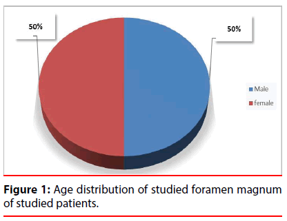
Equal proportion (50%) of cases were males and females (Figure 1).
Table 1 reveals that the youngest aged 18 and eldest was 93 years, median=45 years and mean ± Std. deviation was 46.17 ± 17.80 years.
| Descriptive statistics of age | Values in years |
|---|---|
| Mean | 46.17 |
| Median | 45 |
| Mode | 18 |
| Std. Deviation | 17.8 |
| Minimum | 18 |
| Maximum | 93 |
Table 1: Descriptive statistics of age of foramen magnum of studied patients.
▪ Description of FM
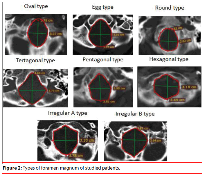
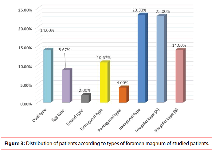
Regarding the shape of FM, there are different shapes of FM were observed which classified into eight types; Oval, Egg, Round, Tetragonal, Pentagonal, Hexagonal type, Irregular type (A) and Irregular type (B) as shown by Figure 2. The most prevalent types hexagonal type (23.33%) and the lowest prevalent type was round (2%). Regarding the proportion of irregular type (A) it was 23%. Equal proportions (14%) of patients had irregular type B and oval as illustrated by Figure 3.
▪ Parameters of FM
Comparative statistics with the maximum and minimum values, means and standard deviations for each dimension of the FM in both males and females are presented in Table 2. Regarding the sagittal diameters of male and female patients; the maximum and minimum values were 46.40 mm and 30.50 mm and 46.00 mm and 26.2 mm respectively. Their means ± St D, were 39.04 ± 3.71 mm and 36.71 ± 3.69 mm respectively.
| Male (n=75) | Female (n=75) | p-value | ||||||||
|---|---|---|---|---|---|---|---|---|---|---|
| Measurements of FM | Mean | SD | Max | Min | Mean | SD | Max | Min | ||
| Sagittal diameter in mm | 39.04 | 3.71 | 46.4 | 30.5 | 36.71 | 3.69 | 46 | 26.2 | 0 | |
| Transverse diameter in mm | 32.4 | 3.11 | 42.2 | 27.1 | 30.76 | 3.22 | 41.6 | 21.9 | 0.002 | |
| FM index | 71.45 | 5.97 | 88.5 | 60.4 | 67.48 | 6.2 | 83.3 | 52.4 | 0 | |
| FM area/T mm2 | 1008.98 | 170.34 | 1537 | 716 | 901.08 | 165.51 | 1362 | 539 | 0 | |
| FM area/R mm2 | 998.13 | 168.32 | 1534 | 716 | 892.3 | 164.67 | 1347 | 533 | 0 | |
Table 2: Minimum, maximum, means, standard deviations and P values of foramen magnum parameters of studied males and females.
These differences were statistically significant (p<0.000).
Concerning, the transverse diameters of male and female patients; The maximum and minimum values were 42.20 mm and 27.10 mm and 41.60 mm and 21.90 mm respectively. Their means ± St D, were 32.40 ± 3.11 mm and 30.76 ± 3.22 mm respectively. These differences were statistically significant (P<0.002).
The male FM index maximum was 88.50 and the minimum was 60.40, these values were greater than females (83.30 and 52.40 respectively). Mean ± St D of males’ FM index (71.45 ± 5.97) was significantly higher than females (67.48 ± 6.20), p<0.000.
According to Teixeira’s formula which was used for calculating the surface area, the maximum and minimum areas of males were 1537 and 716 mm2 and females were 1362 and 539 mm2 respectively. There were statistically significant differences between males’ mean ± StD and females (1008.98 ± 170.34 mm2 and 901.08 ± 165.51 mm2 respectively), p<0.000.
When Ritual’s formula was used for calculating the surface area, similar findings were observed. Males had higher values of maximum and minimum values (1534 and 716 mm2 respectively) compared to females (1347 and 533 mm2 respectively). There were also significant statistical differences between the means ± SD of males and females; (998.13 ± 168.32 and 892.3 ± 164.67 mm2 respectively), (p<0.002).
Discussion
The types and parameters of FM regarding had been studied by many investigators. Based on the findings of our study, the most common types of foramen magnum were both irregular type(A) and hexagonal type. Similarly, Zaidi and Dayal reported that the hexagonal type is the most common type [12]. On the other hand, many studies revealed that ovale type is the most common [13-15]. Murshed et al. [2] and Sharma et al. [15] found out that the most common type was round type. The tetragonal type was reported to be the most common type by Sindel et al. [16]. The difference in most common type of FM could be attributed to different ethnic groups studied by these researches. Murshed et al. and Chethan concluded similar opinion [2,17,18]. The variations in types of FM are important in neurological interpretations in case of ovoid type, surgeons during operative procedures may find difficulty to explore the anterior portion of FM.
The present study revealed that the parameters associated with the FM such as sagittal diameter, transverse diameter, index, and FM area were larger in males compared to females.These findings are in accordance with bulk of studies. Overall, our findings of various parameters were relatively higher than those found in the previously mentioned studies. Concerning with the imaging of FM, the present study detected that the studied Libyan population’s parameters were larger compared to other ethnic groups [2,5,10,18-25].
Furthermore, the present study illustrated that the mean sagittal diameter was 39.04 mm in males and 36.71 in females. The mean transverse diameter was 32.40 mm in males and 30.76 in females. The mean FM index was 71.45 in males and 67.48 in females. The mean FM area/T was 1008.98 mm2 in males and 901.08 mm2 in females. The mean FM area/ R was 998.13 mm2 in males and 892.30 mm2 in females.
Though, it was reported high parameters values such as; the mean sagittal diameter was 37.71 mm in males and 34.37 in females, the mean transverse diameter was 31.68 mm in males and 28.34 in females,the mean FM area/T was 946.66 mm2 in males and 773.96 mm2 in females and the mean FM area/R was 939.47 mm2 in males and 766.81 mm2 in females [14]. Still, our findings are considered higher compared to their findings. It seems that different ethnic groups have differences in the dimensions of the foramen magnum. Another reason for these differences could be related to the different measurement techniques or due to the variety of studied samples.
After puberty, age does not affect the parameters of the foramen magnum. Therefore, differences in the results of various studies could not be due to the age of the samples and there is no need to consider age while determining the parameters of the foramen magnum.
Conclusion
It is concluded that the commonest type of the FM was hexagonally followed by irregular type A. Additionally, our results illustrated that FM parameters were higher in males compared to females. Furthermore, types and parameters of FM should be taken into consideration during radiological reports and during surgical interventions.
Recommendations
Further studies using a larger population size to enable us to generalize the findings for the Libyan population.
References
- Standring S. The anatomical basis of clinical practice: Gray’s anatomy. (39th ed) London Elsevier Churchill Livingstone 23, 492 (2006).
- Murshed KA, Cicekcibasi AE, Tuncer I. Morphometric evaluation of the foramen magnum and variations in its shape: A study on computerised tomographic images of normal adults. Turk J Med Sci 33(5), 301-306 (2003).
- Zaidi SH, Dayal SS. Variations in the shape of foramen magnum in Indian skulls. Anat Anz 167(4), 338-340 (1988).
- Uthman AT, Al-Rawi NH, Al-Timimi JF. Evaluation of foramen magnum in gender determination using helical CT scanning. Dentomaxillofac Radiol 4(3), 197-202 (2012).
- Manoel C, Prado FB, Caria PHF, et al. Morphometric analysis of the foramen magnum in human skulls of brazilian individuals: Its relation to gender. Braz J Morphol Sci 26(2), 104-108 (2009).
- Tubbs RS, Griessenauer CJ, Loukas M, et al. Morphometric analysis of the foramen magnum: An anatomic study. Neurosurgery 66(2), 385-388 (2010).
- Kanchan T, Gupta A, Kewalkrishan K. Craniometric analysis of foramen magnum for estimation of sex. Int J Med Health Biomed Pharma Eng 7(7), 378-380 (2013).
- Suazo I, Russo P, Zavando A, et al. Sexual diamorphism in the foramen magnum dimensions. Int J Morphol 27(1), 21-23 (2009).
- Edward K, Viner M, Schweitzer W, et al. Sex determination from the foramen magnum. J Forensic Radiology Imaging 1(4), 186-192 (2013).
- Burdan F, Szumilo J, Walocha J, et al. Morphology of the foramen magnum in young Eastern European adults. Folia Morphol (Warsz) 71(4), 205-216 (2012).
- Zaidi SH, Dayal SS. Variations in the shape of the foramen magnum in Indian skulls. Anat Anz Jena 167(4), 338-340 (1998).
- Radhakrishnan P, Gupta C, Kumar S, et al. A morphometric analysis of the foramen magnum and variations in its shape: A computerized tomographic study. Novel Science Int J Med Sci 1(9-10), 281-285 (2012).
- Aghakhani K, Kazemzadeh N, Ghafurian F, et al. Gender determination using diagnostic values of foramen magnum. Int J Med Toxicol Forensic Med 6(1), 29-35 (2016).
- Ganapathy A, Sadeesh T, Rao S. Morphometric analysis of foramen magnum in adult human skulls and CT imgages. Int J Cur Res Rev 6(20), 11-15 (2014).
- Sharma S, Sharma A, Modi B, et al. Morphometeric evaluation of the foramen maganum and variation in its shape and size: A study on human dried skull. Int J Anat Res 3(3), 1399-1403 (2015).
- Sindel M, Ozkan O, Ucar Y, et al. Foramen magnum anatomic varyasyonlari. Akd U Tip Fak Dergisi. 6, 97-102 (1989).
- Chethan P, Prakash KG, Murlimanju BV, et al. Morphological analysis and morphometry of the foramen magnum: An anatomical investigation. Turk Neurosurg 22(4), 416-419 (2012).
- Shanthi CH, Lokanadham S. Morphometric study on foramen magnum of human skulls. Medicine Science 2(4), 792-798 (2013).
- Gapert R, Black S, Last J. Sex determination from the foramen magnum: discriminant function analysis in an eighteenth and nineteenth century British sample. Int J Legal Med. 123(1), 25-33 (2009).
- Jain D, Jasuja OP. Evaluation of foramen magnum in sex determination from human cranial by using discriminate function analysis. El Mednifico Journal 2(2), 89-92 (2014).
- Uysal S, Gokharman D, Kacar M, et al. Estimation of sex by 3D CT measurements of the foramen magnum. J Forensic Sci 50(6), 1310-1314 (2005).
- Sendemir E, Savci G, CimenA. Evaluation of the foramen magnum dimensions. Kaibogaku Zasshi 69(1), 50-52 (1994).
- Burdan F, Umlawska W, Dworzanski W, et al. Anatomical variances and dimensions of the superior orbital fissure and foramen ovale in adults. Folia Morphol (Warsz) 70(4), 263-271 (2011).
- Raghavendra YP, Kanchan T, Attiku Y, et al. Sex estimation from foramen magnum dimensions in an Indian population. J Forensic Leg Med 19(3), 162-167 (2012).
- Gapert R, Black S, Last J. Test of age-related variation in the craniometry of the adult human foramen magnum region: implications for sex determination methods. Forensic Sci Med Pathol 9(4), 478-488 (2013)


