FIT OF CAD\CAM FRAMEWORKS DESIGNED BASED ON VIRTUAL IMPLANT POSITIONS VERSUS ACTUAL IMPLANT POSITIONS: A PILOT INVITRO STUDY
- Corresponding Author:
- Mohamed El-Sayed Kamel, Department of Implantology, Faculty of Dentistry, Cairo University, Cairo, Egypt.
E-mail: mohamed.eletr@dentistry.cu.edu.eg
Received: 12-Apr2023, Manuscript No. IJOCS-22-125661; Editor assigned: 17-Apr-2023, PreQC No. IJOCS-22-125661(PQ); Reviewed: 15-Dec-2023, QC No. IJOCS-22-125661(Q); Revised: 25-Dec-2023, Manuscript No. IJOCS-22- 125661(R); Published: 26-Jan2024, DOI: 10.37532/1753- 0431.2024.18(1).243
Abstract
Aim
The aim of this study was to evaluate the fit of CAD\CAM frameworks deigned based on virtual implant positions versus actual implant positions.
Methodology
Five models with 20 frameworks were included. Their cone-beam computerized tomographic images were Planmeca Imaging System and their intraoral surface scans with Medit scanner. The data from these two sources were then merged; the volumetric topography of models was constructed and prosthesis/implant planning was performed using RealGuide software. From these plans, fully computer guided surgical templates and screw-retained Final metal prostheses were manufactured before implant placements using computer–aided design/computer–aided manufacturing process. Jdental implants were placed fully guided, the prostheses were inserted and evaluated based on their time spent for construction. The control group was frameworks done by the conventional technique by scanning the scan bodies of actual implant positions after implant placement.
Results
Results showed that the difference between the two groups was not statistically significant was reported between two groups regarding fit of frameworks
Conclusions
Preoperative frameworks have the same fit as the postoperative frameworks.
Keywords
CAD\CAM, Prefabricated frameworks, Free end saddle, Implants, Guided surgery.
Introduction
Computer assistance in implant dentistry has been accepted in daily practice and is becoming a hot topic in dental implant research. The accuracy of implant position using computerguided templates has been validated clinically and proven superior to free hand implant placement. Systemic reviews, scientific consensus and textbooks on this topic are available [1-4]. Ganz has been studying this field for more than two decades [1-3].
In Free end partial edentulous arches, once implants have been placed fully guided and immediate loading is feasible, there are several methods of fabricating definitive crowns/bridges either directly or indirectly using actual implant positions as the reference. In other words, temporary prostheses made post-operatively involve the physical recording of the implants, their abutments and peri-implant tissues, which are physically manipulated in the fabricating process.
With advances in all aspects of digital techniques, precise preoperative planning for implant surgery and prefabricated implant-supported prosthesis has become fit [5]. Prefabricated prostheses can better achieve esthetic and functional outcomes at the time of surgery [6,7]. Data obtained using Cone-Beam Computerized Tomography (CBCT) can be imported into implant planning software programs to analyze the surrounding vital anatomic structures to determine the ideal implant locations [8]. Intraoral scanning devices help create a more realistic view of the intraoral soft tissues [4]. Optimal prosthetic-driven implant placement can be scheduled virtually before surgery using a scanning template [9]. Digital data from CBCT and intraoral scans can be directly transferred to the manufacturer of surgical templates and final prostheses [8, 10].
This in vitro pilot study aims at exploring the clinical fit of these pre-operative definitive prostheses which are constructed using CBCT without radiographic markers, intraoral scanner, implant planning software and CAD-CAM pathway.
Materials and Methods
■Construction of surgical template
Stone cast of bilateral free end saddle mandibular jaw model was scanned and radiographed using CBCT. The design of the surgical guide was carried out on implant planning software then printed using LCD 3D printer.
■ Drilling of the implants
After checking the seating of the surgical guided stent on the models, drilling was initially performed using drills of diameter size of 2.3 mm (pilot drill), followed by 2.8 mm drills and followed by 3.4 mm then finally 3.8 mm drills for the placement of implants 3.7 mm x10 mm in dimension. The drilling site was cleaned and the fixture installed in place carefully and tightened using contra angled hand piece and a torque wrench.
■Pre-operatively fabricated frameworks: (Intervention group)
This group was restored using pre-operatively fabricated frameworks. The virtual scan bodies of the multi-unit abutments were exported to the designing software, the design of the frameworks were done and then exported for CNC computer aided machining.
■Post-operatively fabricated frameworks: (Control group)
• After the implants installed in the anterior and premolar areas. The multi-unit abutments screwed to the implants. A digital impression using an intra-oral scanner at multi-unit abutment level was carried out by using scan bodies which connected to the six installed implants on the reference cast (control) by hand tightening.
• The digital volumes have been exported as STL files and transferred to the designing software. The design then transferred to the computer aided machine.
■Fit assessment
The fit assessment was binary by applying screw resistance test to the frameworlls.
Results
All data were collected in an excel sheet for statistical analysis. Since they were all qualitative, they were presented as frequencies and percentages. Fisher’s Exact test was used to compare between the two groups. The significance level was set at P ≤ 0.05. Statistical analysis was performed with IBM SPSS Statistics for Windows, Version 23.0. Armonk, NY: IBM Corp (Table 1).
Table 1: Results of Fisher’s Exact test for comparison between overall feasibility of frameworks
| Group | Overall fit | N | % | ||
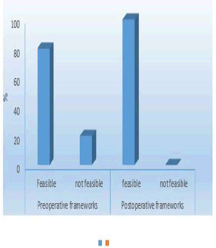
| Group prefabricated | yes | 9 | 90 | 0.1 | NC |
| no | 1 | 10 | |||
| Group Post-operatively | yes | 10 | 100 | NC | NC |
| no | 0 | 0 | |||
| %: percentage, *: Significant at P ≤ 0.0, N: number, NC: not computed because of constant variable, OR: | |||||
■Odds ratio
The results showed that no significant difference between the preoperatively and postoperatively fabricated frameworks regarding fit (Figure 1).
Discussion
This in-vitro study compares the fit between the frameworks fabricated before and after implant placement using complete digital workflow.
The need of pre operatively fabricated frameworks is increasing to save time, cost and effort. Also, it encourages the clinicians to adopt the completer digital workflow which facilitates the fabrication of implant supported frameworks with less errors than the conventional techniques.
The merge between the CBCT and intra oral scanning increase the precision of the outcome of the procedure [9]. The use of guided implant placement with computer aided and manufacturing makes fabrication of prefabricated frameworks more precise [2].
The frameworks that were done postoperatively need digital scans after implant placement while this step was skipped in the other group. The step of taking physical or digital impression will save the time and will be more convenient for patients with gagging reflex or allergy to the impression materials [4].
The postoperatively fabricated frameworks need 10 minutes for scanning step and importing the scan to the designing software while the computer aided machining for both groups was the same.
The preoperatively group need more adjustments for complete seating of the frameworks. While the postoperative group needed less adjustments which saved more time.
The cost needed for the both groups considered to be same except for the scanning step. The scanning step is important to transfer the actual positions of the multiunit of the implants which lead to more accurate frameworks. This reduces the need to adjustments and the time of adjusting the frameworks while for the prefabricated frameworks, they need more adjustments which increase time and cost for production of accurately fabricated frameworks. But in the whole procedure of production and adjusting of prefabricated frameworks takes less time than the postoperatively fabricated frameworks.
Conclusion
Within the limitations of the study, we concluded that the preoperatively fabricated frameworks have same feasibility than conventional fabricated frameworks.
Conflict of Interest
The authors declare no conflict of interest.
Funding
This research received no specific grant from any funding agency in the public, commercial, or not-for-profit sectors
Ethics
This study protocol was approved by the ethical committee of the faculty of dentistry- Cairo university on:26\5\2020
References
- Ganz, S. D. Three-dimensional imaging and guided surgery for dental implants. Dent Clin 59, 265-290 (2015).
- Laleman, I., Bernard, L., Vercruyssen, M., et al. Guided Implant Surgery in the Edentulous Maxilla: A Systematic Review. Int J Oral Maxillofac Implants 31(2016).
- Ma, B., Park, T., Chun, I., et al. The accuracy of a 3D printing surgical guide determined by CBCT and model analysis. J Adv Prosthodont 10,279(2018).
- Richert, R., Goujat, A., Venet, L., et al. (2017). Intraoral scanner technologies: a review to make a successful impression. J Healthc Eng (2017).
- D'haese, J., Ackhurst, J., Wismeijer, D., et al. Current state of the art of computer‐guided implant surgery. Periodontology 2000,73,121-133(2017).
- Lee, C. Y., Ganz, S. D., Wong, N., et al. Use of cone beam computed tomography and a laser intraoral scanner in virtual dental implant surgery: part 1. Implant Dent 21, 265-271(2012).
- Makarov, N., Pompa, G., & Papi, P. Computer-assisted implant placement and full-arch immediate loading with digitally prefabricated provisional prostheses without cast: a prospective pilot cohort study. Int J Implant Dent 7, 1-9(2021).
- Yuzbasioglu, E., Kurt, H., Turunc, R., et al. Comparison of digital and conventional impression techniques: evaluation of patients’ perception, treatment comfort, effectiveness and clinical outcomes. BMC Oral Health 14, 1-7(2014).
- Albiero, A. M., & Benato, R. Computer‐assisted surgery and intraoral welding technique for immediate implant‐supported rehabilitation of the edentulous maxilla: case report and technical description. Int J Med Robot Comput Assist Surg 12, 453-460(2016).
- Chen, C., Lai, H., Zhu, H., et al. Digitally prefabricated versus conventionally fabricated implant-supported full-arch provisional prosthesis: a retrospective cohort study. BMC Oral Health 22, 335(2022).
