Evaluation of a Modified CE Angle Measurement to Evaluate Developmental Dysplasia of the Hip
- Corresponding Author:
- S Hagmann
Clinic for Orthopaedics and Trauma Surgery
Centre for Orthopaedics, Trauma Surgery and Spinal Cord Injury
Heidelberg University Hospital, Schlierbacher Landstrasse, Heidelberg, Germany
E-mail: katharina.gather@med.uni-heidelberg.de
Abstract
ABSTRACT
Background: The center edge-angle of Wiberg became one of the main tools to evaluate the lateral coverage of the femoral head in patient from 4 to 75 years. One limitation is the difficulty to define a reliable and accurate center of the femoral head in younger patients. With the improvement of conservative and surgical techniques in the treatment of DDH children are increasingly younger than 4 years at the time of treatment. Thus, it is necessary to have a reliable method to help decision making and to evaluate therapeutic success at this age.
Materials and Methods: Anterior-posterior pelvic radiographs of 33 children (65 hips) aged 4 to 16 years, that had been presenting with developmental hip dysplasia (39/65) or coxitis fugax (11/65) in our pediatric orthopedics outpatient clinic were analyzed in a retrospective analysis. The CE-angle of Wiberg and the modified CE-angle were measured.
Results and Conclusion: X-rays were analyzed by a student, a resident, and an orthopedic specialist. There was a very strong correlation between all three assessors (p<0.001). The reliability of the CE-angle of Wiberg and the modified CE-angle were both excellent with a Cronbach-alpha of 0.92 and 0.912, respectively. Femoral head coverage is one of the most significant prognostic factors for the development of the hip. Therefore, a radiographic tool valid for very young children needs to be established. The modified CE-angle is promising in this regard.
Keywords
CE angle Wiberg; Developmental hip dysplasia; Femoral head coverage; Modified CE angle
Introduction
Since its description in1939, the center edge (CE) angle of Wiberg is one of the main tools to evaluate the lateral coverage of the femoral head in children and adults [1-4].
Although a lot of studies showed it to be a reliable and reproducible measure, it can be difficult to define the center of the femoral head when its appearance is delayed or eccentrically located [5,6]. In patients with deformity of the femoral head, for instance in congenital dysplasia, avascular necrosis or other diseases, defining the center of the femoral head can be especially difficult [6]. This makes the measurement of the CE-angle of Wiberg in younger patients challenging, resulting in potential unreliability. Similar problems exist with the Tönnis classification, which also depends on the existence and position of the center of the femoral head [5]. The International Hip Dysplasia Institute (IHDI) has developed a new radiographic classification to quantify the severity of the displacement of the femoral head not relying on the presence of the ossified nucleus. This classification, therefore, can be applied to children of all ages [5]. The IHDI uses the midpoint of the epiphyseal growth plate instead of the center of the femoral head. This approach seems applicable to the measurement of the CE-angle of Wiberg as well
In 1939, Wiberg evaluated his CE-angle in patients from 8 to 75 years and extended the range in 1955 to a margin of 4 years [2,7]. One reason for this limitation might be the mentioned difficulty to define a reliable and accurate center of the femoral head in younger patients. For children younger than 4 years the reliability of Acetabular Index (AI) is superior to CE-angle of Wiberg, which is why it is mainly used for evaluation of DDH in children younger than four years. However, the AI gives no information about femoral head coverage, which is crucial to evaluate the need for treatment.
With the improvement of both conservative and surgical treatment for congenital dysplasia of the hip, early detection and thus early treatment using the natural growth potential is vital. Successful treatment, however, relies on reliable and comparable measurements to define and quantify the degree of dysplasia. It is thus fundamental to establish a measuring method for hip dysplasia under the age of 4 that is not purely based on Hilgenreiner’s angle and does not change in relation to the configuration of the femoral head.
Our intention was to compare the interobserver reliability of a modified CE angle measurement, using the midpoint of the epiphyseal growth plate as a reference, with the one of Wiberg. To our knowledge, we are the first to describe this reference point for the CE-angle.
Patients and Methods
▪ Patients
Anterior-posterior pelvic radiographs of 33 children (65 hips) that had been presenting with developmental hip dysplasia (39/65) or coxitis fugax (26/65) in our pediatric orthopedic outpatient clinic were analyzed in a retrospective analysis (Table 1). Included are 15 hips that were asymptomatic and radiographically showing no signs of pathology but had been on an x-ray taken for the pathology of the contralateral side. The average age was 8.7 years (SD=3.7 years), ranging from 4 to 16 years. Most patients were female (63%, 42/67; male 37%, 25/67). Patients younger than 4 years were excluded due to the approved age of measurement of the CE-angle of Wiberg. Patients in which the growth plate of the femur had closed were also excluded.
| n | Age in years | Male/Female | Left/Right | |
|---|---|---|---|---|
| Total | 65 | 8.8 (SD=3,7) | 23/42 | 33/32 |
| DDH | 39 | 9.9 (SD=3,6) | 11/28 | 19/20 |
| Coxitis/normal | 26 | 7.1 (SD=3,2) | 12/14 | 14/12 |
Table 1: Total number, average age (in years), sex ratio and number of left and right hips in total and separated in two subgroups (DDH Developmental Hip Dysplasia, normal hips or coxitis fugax) are displayed.
▪ Methods
Radiographs of children without any previous surgery were analyzed. Measurements were made on anterior-posterior pelvic x-rays. Therefore, patients were lying on their back with a slight internal rotation of the leg so that the big toes touched. Included were patients older than four years. All radiographs were analyzed by a student, a resident, and an orthopedic consultant.
The CE-angle of Wiberg was measured as formerly described [2,7]. Briefly, the angle was defined as the angle between a line from the center of the femoral head to the lateral bony edge of the acetabulum and a perpendicular line through the center of the femoral head. The center of the femoral head was first obtained using a circle that outlined the femoral head. The perpendicular line was adjusted to be rectangular to Hilgenreiner’s line.
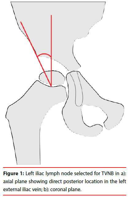
The modified CE-angle was measured in the same way, except that instead of referring to the center of the femoral head, the center of the growth plate was used as a reference (Figure 1).
▪ Statistical analysis
All statistical analysis was conducted using SPSS (version 22; SPSS, Chicago, USA) [8]. Cronbach’s alpha as a measurement for internal consistency. Correlations were calculated with Pearson-Correlation and Spearman-Rho.
Results
65 hips (33 children) were analyzed by a student, a resident, and an orthopedic specialist. The range and means are summarized in Table 2.
| Minimum | Maximum | Mean | SD | ||
|---|---|---|---|---|---|
| CE-angle of wiberg (degree) | |||||
| Student | 2.8 | 34.5 | 18.06 | 7.7 | |
| Resident | 1.1 | 31.3 | 17.9 | 7.5 | |
| Orthopedic specialist | 3.0 | 36.0 | 18.7 | 8.3 | |
| Modified CE-angle (degree) | |||||
| Student | 7.9 | 38.0 | 23.3 | 8.4 | |
| Resident | 4.6 | 37.2 | 20.6 | 7.8 | |
| Orthopedic specialist | 2.0 | 38.0 | 20.0 | 8.6 | |
Table 2: Results for the measurements of the modified CE-angle and the CE-angle of Wiberg (student, resident, consultant).
The reliability of the CE-angle of Wiberg and the modified CE-angle were both good with a Cronbach-alpha 0.92 and 0.91. With a significance of p<0.001 the results of the student did not differ from the orthopedic specialist for both CE-angles. There was a very strong correlation between all three assessors (p<0.001). Pearson-correlation was 0.74-0.79 for the modified CE- angle and 0.77-0.85 for the CEangle of Wiberg, meaning there is a positive connection between the measured CE-anglevalue and all three raters. The same correlation could be observed for Spearman-Rho (0.74-0.78 for the modified CE-angle and 0.755-0.838 for the CE-angle of Wiberg). The inter-itemcorrelation- coefficient was 0.75-0.79 for the modified CE-angle and 0.77-0.85 for the CEangle of Wiberg.
All patients were divided into four age subgroups. The first group ranged from 4 to 6 years, the second from 7 to 9, the third from 10 to 13 and the forth from 14-16 years. The results of the measurements were compared as described above for all assessors. For both CE-angles, no significant difference in the measurements was observed between the age groups.
Discussion and Conclusion
Due to the improvement of early detection of developmental hip dysplasia with ultrasonic hip screening and with the improvement of the surgical techniques for treating the disease, patients are often much younger than 4 years at the time of surgery. These patients are not limited to those needing open reduction for displacement but also comply with patients undergoing Dega acetabuloplasty or Salter osteotomy in combination with an osteotomy of the proximal femur. Recent studies show an average age at time of surgery from 1.8 to 4.5 years [9-12].
Radiographs are mainly used as the basis for treatment decisions after the children have reached walking age. Often enough, treatment decisions are based on criteria such as acetabular configuration or coverage of the femoral head. While Hilgenreiner‘s index is commonly used to evaluate the acetabular configuration, the CEangle of Wiberg, as one of the best diagnostic tools to quantify coverage of the femoral head is not verified for a young age. Even the reliability of the acetabular index, mainly used in this age, is lower for patients under 3 years [13]. The need for a measurement tool under the age of 4 and the limits of current values have also shown by other authors [14,15].
In addition, there is not only a need to quantify treatment decision criteria, hopefully enabling a more scientific and reproducible approach for surgical treatment in DDH, but also a need to quantify the results of surgery. The acetabular index is useful for the evaluation of acetabular osteotomies in this regard, however, restoration of femoral head coverage cannot be objectified, although being one of the main reasons for surgery. Using a modified CE-angle for children of young age is our address to this problem in the future. We therefore aimed at demonstrating that the modified CE-angle is a tool with similar accuracy in the validated range and to show that the modified CE-angle is comparable to the classical CE-angle of Wiberg regarding its reliability. Three raters with different levels of training showed no difference in reliability between the modified CE-angle and the one of Wiberg. Therefore, the modified CE-angle may prove useful as an alternative in children of young age or for other reasons when the classical CE-angle may not be applicable.
Our reliability results for both angles are comparable with other publications concerning the CE-angle of Wiberg [4,16-18]. We believe that when measuring a larger number of patients, it may prove the easier way to locate the reference center of the angle with the middle of the growth plate, basically because it is more referencing the center of a line than referencing the center of an oval or circle. This could be especially useful in children younger than 4 years, where the CEangle of Wiberg has already been proven to be unreliable [2,7].
Concerns using the modified CE-angle may include the variance in the shape of the epiphyseal plate at different ages. However, in our study, intra- and interobserver reliability did not change in the different age groups. Also, the femoral head itself shows important variations depending on the age of the children.
The aim of this study was to evaluate whether a modified CE-angle had comparable interand intraobserver reliability. In our opinion, this is crucial before beginning to evaluate the differences between the two angles and to define thresholds. The practical need for quantification of femoral head coverage in very young children calls for further evaluation of this modification.
Femoral head coverage is the most significant prognostic factor for the development of the hip in DDH and an important way of deciding on treatment and evaluating its success. Therefore, a radiographic tool valid for very young children to quantify femoral head coverage needs to be established. Showing comparable reliability, the modified CE-angle is promising in this regard.
Conflict of Interest
The authors declare that they have no conflict of interest.
References
- Engelhardt P. The significance of the center-edge angle in the prognosis of the dislocated hip 50 years after its initial description by Wiberg. Orthopade 17(6), 463-467 (1988).
- Fredensborg N. The CE angle of normal hips. Acta Orthop Scand 47(4), 403-405 (1976).
- Wientroub S, Tardiman R, Green I, et al. The development of the normal infantile hip as expressed by radiological measurements. Int Orthop 4(4), 239-241 (1981).
- Nelitz M, Guenther KP, Gunkel S, et al. Reliability of radiological measurements in the assessment of hip dysplasia in adults. Br J Radiol 72(856), 331-334 (1999).
- Narayanan U, Mulpuri K, Sankar WN, et al. Reliability of a new radiographic classification for developmental dysplasia of the hip. J Pediatr Orthop 35(5), 478-484 (2015).
- Tonnis D. Normal values of the hip joint for the evaluation of X-rays in children and adults. Clin Orthop Relat Res (119), 39-47 (1976).
- Wiberg G, Jerre T. Importance of early diagnosis of epiphysiolysis of the hip. Sven Lakartidn 52(23), 1496-1501 (1955).
- IBM Corp. IBM SPSS Statistics for Windows, Version 22.0. Armonk, IBM Corp, NY (2013).
- El-Sayed M, Ahmed T, Fathy S, et al. The effect of dega acetabuloplasty and Salter innominate osteotomy on acetabular remodeling monitored by the acetabular index in walking DDH patients between 2 and 6 years of age: Short- to middle-term follow-up. J Child Orthop 6(6), 471-477 (2012).
- Ming-Hua D, Rui-Jiang X, Wen-Chao L. The high osteotomy cut of dega procedure for developmental dysplasia of the hip in children under 6 years of age. Orthopade 45(12), 1050-1057 (2016).
- Ramani N, Patil MS, Mahna M. Outcome of surgical management of developmental dysplasia of hip in children between 18 and 24 months. Indian J Orthop 48(5), 458-462 (2014).
- Akgul T, Bora Goksan S, Bilgili F. Radiological results of modified dega osteotomy in tonnis grade 3 and 4 developmental dysplasia of the hip. J Pediatr Orthop B 23(4), 333-338 (2014).
- Upasani VV, Bomar JD, Parikh G. Reliability of plain radiographic parameters for developmental dysplasia of the hip in children. J Child Orthop 6(3), 173-176 (2012).
- Firth GB, Robertson AJ, Ramguthy Y, et al. Prognostication in developmental dysplasia of the hip using the ossific nucleus center edge angle. J Pediatr Orthop 38(5), 260-265 (2018).
- Kitoh H, Kitakoji T, Katoh M, et al. Prediction of acetabular development after closed reduction by overhead traction in developmental dysplasia of the hip. J Orthop Sci 11(5), 473-477 (2006).
- Broughton NS, Brougham DI, Cole WG, et al. Reliability of radiological measurements in the assessment of the child's hip. J Bone Joint Surg Br 71(1), 6-8 (1989).
- Tannast M, Mistry S, Steppacher SD, et al. Radiographic analysis of femoroacetabular impingement with hip2norm-reliable and validated. J Orthop Res 26(9), 1199-1205 (2008).
- Mast NH, Impellizzeri F, Keller S, et al. Reliability and agreement of measures used in radiographic evaluation of the adult hip. Clin Orthop Relat Res 469(1), 188-199.
