Effect of Modified Reverse-6 Taping Procedure with Elastic Tape on Medial Longitudinal Arch in Patients with Patellofemoral Pain Syndrome
- Corresponding Author:
- D. Rajesh
Assistant professor, SRM College of Department of Physiotherapy
SRM University, India
Tel: +91-44-43969999, 43969975
Fax: +91-44-23624778
Email: drajeshdinakaran@gmail.com
Abstract
Objective: The purpose of this study was to find out the effect of “modified Reverse-6 taping procedure” using elastic tape in patellofemoral pain syndrome.
Methodology: Totally 30 participants were included, based on inclusion criteria with age ranges from 18-35 years, height of arch 5.46cm and below, Arch height 5.46 cm and below, Calcaneal eversion 3.8 degree and above, Anterior knee pain scale (KUJALA’S scoring scale ) scored less than 85/100, from various settings like SRM Medical College Hospital and Research Centre (Out Patient – Physiotherapy Department) Kattankulathur, London Ortho Specialty Hospital, Salem and Mahabarathy College of Engineering, Chinna Salem. Pre-test measurements were recorded by using KUJALA’S scale for patellofemoral pain syndrome; arch height and calcaneal eversion were measured before intervention.
Procedure: Four strips of tape- two for modified reverse-6 procedures and another two for heel lock is applied and post-test values were measured at the end of third week.
Results: suggests that the intervention is significantly difference (p <0.05) in improving the Medial longitudinal arch height, reducing calcaneal eversion and in improving KUJALA’S score.
Conclusion: The study concluded that a three week Intervention of modified reverse-6 taping procedure has effect on patellofemoral pain syndrome.
Keywords
Modified reverse-6 taping; Patellofemoral pain syndrome; Medial longitudinal arch
Introduction
Patellofemoral pain syndrome (PFPS) is one of the most common orthopedic conditions [1-3], particularly in young females [3,4]. Murray et al. reported that Patellofemoral pain syndrome accounted for 34% of knee injuries and 10% of all injuries among individuals required treatment at a sports injury clinic. Due to the high recurrence rate of Patellofemoral pain syndrome and a potential associated with the future development of patellofemoral osteoarthritis, this condition warrants further study [5,6].
The etiology of Patellofemoral pain syndrome is likely related to abnormal patellar alignment relative to the femoral trochlea during weight bearing activities and elevated patellofemoral joint stress [7-10]. Altered foot posture and foot and ankle kinematics may contribute to hip and knee joint mechanics thought to increase patellofemoral joint stress [9-11]. Specifically, increased foot pronation has been suspected by these authors to contribute to increased compensatory femoral internal rotation, hip adduction, and knee abduction, resulting in decreased patellofemoral contact area and increased retropatellar stress [9,10].
In addition, individuals with Patellofemoral pain syndrome have been reported to display a greater calcaneal valgus posture and increased navicular drop than subjects without Patellofemoral pain syndrome [12,13] as well as greater hip internal rotation, hip adduction, and knee external rotation during running [14-17]. Additionally, individuals with greater foot mobility during weight bearing appear twice as likely to develop Patellofemoral pain syndrome [18].
Antipronationtaping (APT) is frequently used by clinicians in the management of lowerextremity musculoskeletal pain and injury. Several studies support this practice, reporting that Antipronation taping reduced pain scores in individuals with heel and foot pain immediately after application [19,20], during an intervention period of 1-5 days [19-21]. These studies hypothesized that the reduction of pain was attributable to mechanical changes induced by Antipronation taping, such as alteration in forefoot pressures, navicular height, calcaneal angle, and tibial torsion [19,20]. In support of this hypothesis, several studies have reported mechanical changes induced by Antipronation taping, including increased navicular height, decreased calcaneal eversion, and decreased internal tibial rotation in both resting standing posture [22- 25] and during walking and running [26-28].
Although several different taping techniques have been described to limit foot pronation, the “lowdye” and “Reverse-6” have received the greatest attention. The low-dye technique was described by Dr. Ralph Dye and has shown to increase the height of the medial longitudinal arch [26-29] and also provide short-term relief of symptom associated with plantar fasciitis.[21,27,30,31] The Reverse-6 taping technique has also been used to control excess foot pronation.[24,32,33]. In a recent systematic review, Cheung et al. [34] reported that while adhesive taping was more effective than footwear modification and foot orthoses in controlling foot pronation, the low-Dye technique was less effective than other taping techniques such as the Reverse-6 that is applied above the talocrural joint.
Although control of foot pronation is more effective when the tape is applied above the talocrural joint, the Reverse-6 technique that was originally described by Vicenzino et al. covered both malleoli [24]. Previous research has indicated that talocrural joint range of motion, especially plantar flexion, may be restricted too much when tape is applied over the malleoli [35].
Inelastic tape has been used almost exclusively in order to control foot pronation and restrict foot movement [22,26,30,36]. Only one study has been published in which elastic tape was used to restrict motion of the medial longitudinal arch. In a case series study the authors reported that the use of a “modified” Reverse-6 taping technique using elastic tape reduced symptom associated with foot pronation for a variety of foot and lower extremity disorders [22].
In addition, the change in foot posture created by the “modified” Reverse-6 taping technique could also be used to determine the degree of orthotic posting [33]. In that study, the original Reverse-6 taping technique described by Vicenzino et al. [24] was modified by first altering the path of the tape so that it did not cross the medial or lateral malleoli and therefore would not cause any reduction in talocrural joint range of motion and second by using elastic rather than inelastic tape [33].
While the Meier et al. case study series demonstrated that the “modified” Reverse-6 (MR6) taping technique was clinically effective, it is still critical to understand if it can be reliably applied in order to produce a consistent change in the height of the medial longitudinal arch from one session to the next [33] from one day to the next, and from one clinician to the next. Thus, the purpose of this study was to determine whether the “modified Reverse-6 taping procedure” using elastic tape on medial longitudinal arch in turn to reduce pain in patients with patellofemoral pain syndrome. Reduced medial longitudinal arch is common among individual [37] and the modified reverse-6 taping technique using elastic tape within the same day has immediate effect in the height of the medial longitudinal arch, and has high degree of reliability in physiotherapy practice, hence to explore the long term effect of applying modified reverse-6 taping on medial longitudinal arch [38] calcaneal eversion and pain on patients with patellofemoral pain syndrome. The purpose of the study to find the effect of modified reverse 6 taping technique with elastic tape on medial longitudinal arch in patients with patellofemoral pain syndrome (Figures 1 and 2).
Methodology
Research Design-Quasi Experimental Study, Research Type-Pre Test-Post Test, Sampling Method-Convenient Sampling Sample Size-30 Subjects, Research Setting-SRM Medical College Hospital and Research Centre (Out Patient – Physiotherapy Department) Kattankulathur. London Ortho Specialty Hospital, Salem, Mahabarathy College of Engineering, Chinna Salem Study Duration-3 Weeks.
Procedure
The participants in this study were included from various settings like SRM Medical College Hospital and Research Centre (Out Patient – Physiotherapy Department) Kattankulathur, London Ortho Specialty Hospital, Salem and Mahabarathy College of Engineering, Chinna Salem. Totally 30 participants were included, based on inclusion criteria with age ranges from 18-35 years, height of arch 5.46cm and below, Arch height 5.46 cm and below, Calcaneal eversion 3.8 degree and above, Anterior knee pain scale (KUJALA’S scoring scale ) scored less than 85/100, Subjects those who are willing to participate. Exclusion Criteria: Pregnancy, Foot or ankle injury in one year duration, Traumatic injury in lower limb and Patient undergoing any treatment, and the study was explained in detail in detail, informed consent was obtained.
Measurement Tools
1. KUJALA’S scoring scale
2. Caliper for measuring navicular height
3. Universal Goniometer
Test Procedure
Pretest measurements were recorded by using KUJALA’S scale for patellofemoral Pain subjectively, Arch height measured using caliper and Calcaneal eversion measured using universal goniometer, before initiation of first session and Post-test values were measured at the end of 3rd week respectively.
Tape Application
▪ Pre-taping procedure
Before application of tape, the surface of the foot should clean by isopropyl alcohol, After cleaning applied under wrap over the foot (from fore foot to above the medial malleolus).
The elastic tape measured is taken at appropriate length for subject’s foot, and then the tape is made into four pieces (two pieces is for heel locking and another two for modified reverse-6 procedures) [39].
▪ Modified reverse -6 procedure
The modified reverse-6 taping procedure using elastic tape
1. First strip of tape applied starting on the lateral aspect of the foot and ending above the medial malleolus.
2. The second strip is applied as a reinforcement same as the first trip procedure.
3. The other two strips are used for heel lockbringing everted calcaneum to neutral position; the taping is done starting from the lateral aspect of ankle crossing over to the medial aspect and finally crossing through the posterior calcaneum and ending above the foot towards the lateral aspect of foot [40].
Data Analysis
The statistical package for IBM Compactable (SPSS) version 20 for windows was used for data analysis. The statistical tool used in this study was paired t-test. Paired t-test was used for analysis of pre and post-test means within the group. The collected data were tabulated and analyzed using descriptive and inferential statistics [41], using statistical package for social science (SPSS) version 17.
Results
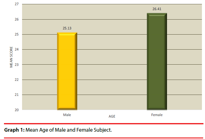
In Graph 1: Shows the distribution of anterior knee subjects according to gender and age. A majority 73.3% of them were females and the rest 26.7% were males. The mean age of males and females was found to be more or less same.
In Table 1 and Graph 2: Presents the statistical outcomes of range, mean and SD of pre and post test of KUJALA’S scoring, before intervention the patellofemoral pain syndrome subjects with mean 73.20 and SD of 4.88 and after the intervention the KUJALA’S scoring was found to be increased to the mean 92.13 with SD of 4.43, and it was found to be statistically significant (P <0.05).
| S. no | AKPS | Range | Mean | SD | Mean difference | Paired t-value | p-value |
|---|---|---|---|---|---|---|---|
| 1 | Pre test | 64-81 | 73.2 | 4.88 | 18.93 | 22.28* | P<0.001 |
| 2 | Post test | 80-98 | 92.13 | 4.43 |
Table 1: Range, mean, SD and paired t-test analysis of KUJALA’S scoring scale among the patellofemoral pain syndrome subjects.
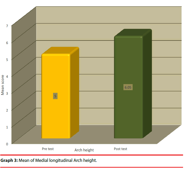
In Graph 3: Depicts the statistical outcomes of mean and SD of pre and post test of navicular height. With mean 5.00 and SD of .131and after the intervention the navicular height was found to be increased to the mean 6.05 with SD of 0.66, and it was found to be statistically significant (P < 0.05).
In Graph 4: Presents the statistical outcomes of mean and SD of pre and post test of Calcaneal eversion. Before the mean was 5.60 and SD of 1.52, and after the intervention the Calcaneal eversion was found to be decreased to the mean 3.63 with SD of 0.92, and again it was found to be statistically significant (P<0.05).
Discussion
This study determined the effects of modified reverse-6 taping procedure with elastic tape on medial longitudinal arch in patients with patellofemoral pain syndrome [42]. PFPS individuals reported to have pronated foot type and increased foot pronation is associated with rare foot eversion [43]. Modified Reverse 6 taping procedure had immediate effects in improving the KUJALA’S scoring among patella femoral pain syndrome subjects and there are several studies supporting the hypothesis. In this study, there was an increase, in navicular height, after the intervention, and it was supported by Landis and Koch [44] study proved regardless of the level of experience modified reverse-6 taping produced change in height and width of medial longitudinal arch, and also by Cornwall, Lebec, Degeyter and McPoil [45] have concluded that there was considerable increase in arch height and width of the midfoot from 62.7 and 78.9mm, to 66.6 and 78.8mm before and after the intervention respectively. However previous studies reported traditional modified reverse 6 and low dye taping using inelastic tape also caused reduction in mid foot width. In this study, after the intervention the Calcaneal eversion was found to be decreased, apparently this study using elastic tape with modified reverse 6 procedures had effects on decrease in calcaneal eversion with mean difference of 1.97cm, an increase in arch height of 1.05 cm. The decrease in calcaneal eversion and increase in medial longitudinal arch indicate that elastic tape using modified reverse 6 procedure is able to alter the foot mechanics during weight bearing. This was supported by Aishwarya and Venkata Sai [46] found that between-groups analysis showed significant reduction of calcaneal eversion angle in calcaneal taping group 3.4 (95% CI = -4.52 to -2.42 ̊) than windlass taping group. This current study using modified reverse 6 taping technique with elastic tape is a standard technique and the type of tape used in previously published studies reference [26,24,28]. Individuals with patella femoral pain syndrome have been reported to demonstrate a pronated foot type [12]. Increased foot pronation may be associated with more rapid rear foot eversion during walking, but only among those with patella femoral pain syndrome [47]. There does not appear to be a generally accepted clinical method to evaluate standing foot posture and different measures were used to classify foot type, foot posture index or the arch height index may be comprehensive and reliable measures of standing foot posture compared with clinical angle [47,48]. However, many static measures of foot pronation have been found to have inconsistent relationships with dynamic foot pronation [49-52].
Decrease hip internal rotation may be particularly beneficial for the treatment of patella femoral pain syndrome since increased hip internal rotation may increase patellofemoral contact area during weight bearing activities [52] further increased hip internal rotation and foot pronation have been identified among females with PFPS, supporting an intuitive link between increased foot pronation and the etiology or exacerbation of PFPS [12,15 ]. This study was tried to found the relationship of mechanics of foot on anterior knee pain that was one of the main strength. Limitations of this study were small sample size, Unequal distribution of gender, no control group. Future studies recommends with larger population size, BMI can be added as a baseline and with equal gender distribution, Specific exercises programs can be added with taping protocol, Baseline assessment with standardized scale e.g. FPI can be used. Future research need to be performed to address the change in width of the mid foot after application of reverse 6 taping.
Future research need to be performed to address the change in width of the midfoot after application of modified reverse 6 taping. In addition previous studies have looked on how long tape will effect a change in foot posture or mobility have used inelastic tape and either low dye or the ‘augmented low due technique, Low dye technique has reported a loss of 20 percent in the height of the navicular bone following a bout of exercise [47]. Subsequently another different study has proved anti pronation taping along with temporary orthotics has effect to control excessive foot pronation and reduces PTPS [12] Antipronation tape application reducing activity of Tibialis Anterior and Tibialis posterior muscles can also alter arch height [49]. Most studies supporting elastic or dye taping have used smaller population and it is recommended from this study that larger population and inter-testing and intra – testing for longer periods can have highly significant results in the management of PFPS. This study concluded that modified reverse-6 taping procedure with elastic tape in patients with patella femoral pain syndrome has brought changes in increasing arch height, reducing Calcaneal eversion and reducing patella femoral pain, and that there is anecdotal evidence that individuals prefer the elastic tape to the inelastic tape because it is more comfortable and that there is less skin irritation and blister formation.
Conclusion
The study concluded that a three week Intervention of modified reverse-6 taping procedure is increased the medial longitudinal arch height, and reduced Calcaneal eversion that in turn decreased the patellofemoral pain syndrome.
References
- Grady EP, Carpenter MT, Koenig CD, et al. Rheumatic findings in gulf war veterans. Arch. Intern. Med 158(4), 367-371 (1998).
- Murray IR, Murray SA, Mackenzie K, et al. How evidence based is the management of two common sports injuries in a sports injury clinic? Br. J. Sports Med 39(12), 912-916 (2005).
- Taunton JE, Ryan MB, Clement DB, et al. A retrospective case-control analysis of 2002 running injuries. Br. J. Sports Med 36(2), 95-101 (2002).
- Fulkerson JP, Arendt EA. Anterior knee pain in females. Clin. Orthop. Relat. Res (372), 69-73 (2000).
- Stathopulu E, Baildam E. Anterior knee pain: A long-term follow-up. Rheumatology (Oxford) 42(2), 380-382 (2003).
- Utting MR, Davies G, Newman JH. Is anterior knee pain a predisposing factor to patellofemoral osteoarthritis. Knee 12(5), 362-365 (2005).
- Doucette SA, Goble EM. The effect of exercise on patellar tracking in lateral patellar compression syndrome. Am. J. Sports Med 20(4), 434-440 (1992).
- Goodfellow J, Hungerford S, Woods C. Patello-femoral joint mechanics and pathology. J. Bone Joint Surg. Br 58(3), 291-299 (1976).
- Powers CM. The influences of altered lower-extremity kinematics on patellofemoral joint dysfunction: a theoretical perspective. J. Orthop. Sports Phys. Ther 33(11), 639-646 (2003).
- Sanchis-Alfonso V, Rosello-Sastre E, Martinez-Sanjuan V. Pathogenesis of anterior knee pain syndrome and functional patellofemoral instability in the active young. Am. J. Knee Surg 12(1), 29-40 (1999).
- Tiberio D. The effect of excessive subtalar joint pronation on patellofemoral joint mechanics: a theoretical model. J. Orthop. Sports Phys. Ther 9(4), 160-169 (1987).
- Barton CJ, Bonanno D, Levinger P, et al. Foot and ankle characteristics in patellofemoral pain syndrome: A case control and reliability study. J. Orthop. Sports Phys. Ther 40(5), 286-296 (2010).
- Levinger P, Gilleard WL. An evaluation of the rearfoot posture in individuals with patellofemoral pain syndrome. J. Sports Sci. Med 3, 8-14 (2004).
- Dierks TA, Manal KT, Hamill J, et al. Proximal and distal influences on hip and knee kinematics in runners with patellofemoral pain during a prolonged run. J. Orthop. Sports Phys. Ther 38(8), 448-456 (2008).
- Souza RB, Powers CM. Differences in hip kinematics, muscle strength, and muscle activation between subjects with and without patellofemoral pain. J. Orthop. Sports Phys. Ther 39(1), 12-19 (2009).
- Willson JD, Davis IS. Lower extremity mechanics of females with and without patellofemoral pain across activities with progressively greater task demands. Clin. Biomech (Bristol, Avon). 23(2), 203-211 (2007-2008).
- Willson JD, Davis IS. Lower extremity strength and mechanics during jumping in women with patellofemoral pain. J. Sport Rehabi 18(1), 76-90 (2009).
- Boling MC, Padua DA, Marshall SW, et al. A prospective investigation of biomechanical risk factors for patellofemoral pain syndrome. Am. J. Sports Med 37(11), 2108-2116 (2009).
- Jamali B, Walker M, Hoke B, et al. Windlass taping technique for symptomatic relief of plantar fasciitis. J. Sport Rehabi 13(3), 228–43 (2004).
- Saxelby J, Betts RP, Bygrave CJ. “Low-Dye’’ taping on the foot in the management of plantar-fasciitis. Foot 7(4), 205-209 (1997).
- Landorf KB, Radford JA, Keenan AM, et al. Effectiveness of low-Dye taping for the short-term management of plantar fasciitis. J. Am. Podiatr. Med. Assoc 95(6), 525-530 (2005).
- Hadley A, Griffiths S, Griffiths L et al. Antipronation taping and temporary orthoses-effects on tibial rotation position after exercise. J. Am. Podiatr. Med. Assoc 89(3), 118 (1999).
- Harradine P, Herrington L, Wright R. The effect of low dye taping upon rearfoot motion and position before and after exercise. Foot 11(2), 57–60 (2001).
- Vicenzino B, Feilding J, Howard R et al. An investigation of the anti-pronation effect of two taping methods after application and exercise. Gait Posture 5(1), 1-5 (1997).
- Vicenzino B, Griffiths SR, Griffiths LA, et al. Effect of anti-pronation tape and temporary orthotic on vertical navicular height before and after exercise. J. Orthop. Sports Phys. Ther 30(6), 333-339 (2000).
- Franettovich M, Chapman A, Blanch P, et al. A physiological and psychological basis for anti-pronation taping from a critical review of the literature. Sports Med 38(8), 617-31 (2008).
- Keenan AM, Tanner CM. The effect of high-dye and low-dye taping on rear foot motion. J. Am. Podiatr. Med. Assoc 91(5), 255-261 (2001).
- Vicenzino B, Franettovich M, Mcpoil T, et al. Initial effects of anti-pronation tape on the medial longitudinal arch during walking and running. Br. J. Sports Med 39(12), 939-943 (2005).
- Vicenzino B, McPoil TG, Russell T, et al. Antipronation tape changes foot posture but not plantar ground contact during gait. Foot 16(2), 91-97 (2006).
- Abd EI Salam MS, Abd Elhafz YN. Low-dye taping versus medial arch support in managing pain and pain-related disability in patients with plantar fasciitis. Foot Ankle Spec 4(2), 86-91 (2011)
- Radford JA, Burns J, Buchbinder R, et al. The effect of low-Dye taping on kinematic, kinetic, and electromyographic variables: a systematic review. J. Orthop. Sports Phys. Ther 36(4), 232-241 (2006).
- Moss CL, Gorton B, Deters S. A comparison of prescribed rigid orthotic devices and athletic taping support used to modify pronation in runners. J. Sport Rehabi 2, 179-186 (1993).
- Meier K, McPoil TG, Cornwall MW, et al. Use of antipronation taping to determine foot orthoses prescription: a case series. Res. Sports Med 16(257), 257-271 (2008).
- Cheung RT, Chung RC, Ng GY. Efficacies of different external controls for excessive foot pronation: a meta-analysis. Br. J. Sports Med 45(9), 743-751 (2011).
- Delahunt E, O’Driscoll J, Moran K. Effects of taping and exercise on ankle joint movement in subjects with chronic ankle instability: A preliminary investigation. Arch. Phys. Med. Rehabil 90(8), 1418–1422 (2009).
- Yoho RR, Rivera JJ, Renschler RR, et al. (2013) A biomechanical analysis of the effects of low-dye taping on arch deformation during gait. Foot 22(4), 283-286 (2013).
- Williams DS, McClay IS. Measurements used to characterize the foot and the medial longitudinal arch: Reliability and validity. Phys. Ther 80(9), 864-871 (2000)
- Vicenzino B, Griffiths SR, Griffiths LA, et al. Effect of antipronation tape and temporary orthotic on vertical navicularhright before and after exercise. J. Orthop. Sports Phys. Ther 30(6), 333-339 (2000).
- Kelly LA, Racinais S, Tanner CM, et al. Augmented low dye taping changes muscle activation patterns and plantar pressure during treadmill running. J. Orthop. Sports Phys. Ther 40(10), 648-655 (2010).
- Ator R, Gunn K, McPoil T, et al. The effect of adhersive straping on medial longitudinal arch support before and after exercise. J. Orthop. Sports Phys. Ther 14(1), 18-23 (1991).
- Lange B, Chipchase L, Evans A et al. The effect of low-Dye taping on plantar pressures, during gait, in subjects with navicular drop exceeding 10 mm J. Orthop. Sports Phys. Ther 34(4), 201-209 (2004).
- Holmes CF, Wilcox D, Fletcher JP. Effect of a modified, low-dye medial longitudinal arch taping procedure on the subtalar joint neutral position before and after light exercise. J. Orthop. Sports Phys. Ther 32(5), 194-201 (2002).
- Picciano AM, Rowlands MS, Worrell T. Reliablity of open and close kinetic chain subtalar neutral position and navicular drop test. J. Orthop. Sports Phys. Ther 18(4), 553-558 (1993).
- Landis JR, Koch GG. The measurement of observer agreement for categorical data. Biometrics 33(1), 159-174 (1977).
- Cornwall MW, Lebec M, Degeyter J, et al. The reliability of the modified reverse-6 taping procedure with elastic tape to alter the height and width of the medial longitudinal arch. Int. J. Sports Phys. Ther 8 (4), 381-392 (2013).
- NC Aishwarya, Sai KV. Immediate Effect of Calcaneal Taping Versus Windlass Taping on Calcaneal Angle in Subjects with Plantar Fasciitis. Int. J. Ther. Appl 33, 28-32 (2016).
- Barton CJ, Levinger P, Crossley KM, et al. Relationships between the foot posture index and foot kinematics during gait in individuals with and without patellofemoral pain syndrome. J. Foot Ankle Res 4, 10 (2011).
- Butler RJ, Hillstrom H, Song J, et al. Arch height index measurement system: Establishment of reliability and normative values. J. Am. Podiatr. Med. Assoc 98(2), 102-106 (2008).
- Levinger P, Gilleard W. Relationship between static posture and rearfoot motion during walking in patellofemoral pain syndrome. J. Am. Podiatr. Med. Assoc 96(4), 323-329 (2006).
- McPoil TG, Cornwall MW. Relationship between three static angles of the rearfoot and the pattern of rearfoot motion during walking. J. Orthop. Sports Phys. Ther 23(6), 370-375 (1996).
- Razeghi M, Batt ME. Foot type classification: a critical review of current methods. Gait Posture 15(3), 282-291 (2002).
- Salsich GB, Perman WH. Patellofemoral joint contact area is influenced by tibiofemoral rotation alignment in individuals who have patellofemoral pain. J. Orthop. Sports Phys. Ther 37(9), 521-528 (2007).


