A Comparative Study to Analyze the Effect of Ultrasound Therapy versus Microcurrent Therapy in Patients with Chronic Lateral Epicondylitis: A Quasi Experimental Study
- Corresponding Author:
- K. Jothi Prasanna
Assistant Professor, College of Physiotherapy Kattankulathur campus
SRM University SRM Nagar
Kattankulathur-603203, Kancheepuram District, Tamil Nadu, India
Tel: 044-47432720; 044-2745-6729
email: jothiprasanna.k@ktr.srmuniv.ac.in
Abstract
Background: Lateral epicondylitis is a relatively common musculoskeletal condition that can cause significant pain and disability. Treatment of lateral epicondylitis aims at reducing pain, increasing grip strength and improving the quality of life of the patient. Therapeutic ultrasound, phonophoresis, LASER therapy, manipulation, soft tissue mobilisation, neural tension, stretching and strengthening has long played an important role in the treatment of lateral epicondylitis, however treatment involving Microcurrent therapy for management of lateral epicondylitis are limited to this date.
Objective: To compare the effectiveness of Ultrasound therapy and Microcurrent therapy in subjects with chronic lateral epicondylitis. Study design: Quasi experimental study design.
Subjects: 20 subjects,10 each with Ultrasound therapy and Microcurrent therapy age group between 35-50 years of both male and female.
Intervention: 10 subjects in group A received Ultrasound therapy with Pre and Post-test and 10 subjects in group B received Microcurrent therapy with Pre and post-test. Outcome measure: Visual Analogue Scale [VAS], Grip Strength and PRTEE.
Results: Statistical analysis was done by using paired ‘t’ test which showed significant improvement in both groups.
Conclusion: Microcurrent therapy has shown significant result in reduction of pain, increased grip strength and functional activity in patients with chronic lateral epicondylitis.
Keywords
Microcurrent, Grip strength, Ultrasound
Introduction
The elbow is a complex hinge joint designed to withstand a wide range of dynamic exertional forces. The elbow is primarily a hinged joint, but possesses the unique ability to rotate the distal arm in pronation and supination [1]. These motions, along with a wide range of dynamic exertional forces, predispose the elbow to various injuries, particularly with repetitive motions.
This overuse tendinopathy occurs in approximately 1% to 3% of the population annually, and although it is commonly called tennis elbow, only 5% to 10% of tennis players develop the condition. Most patients are in their 30s and 40s and develop lateral epicondylitis as a result of occupational rather than recreational activities [2].
The lateral elbow is affected four to 10 times more often than the medial side [3]. The lateral epicondyle of humerus serves as the common extensor origin for the active supinators of the forearm, including the extensor carpi radialis brevis. Physical examination reveals maximal tenderness approximately 1 cm distal to the epicondyle at the origin of the extensor carpi radialis brevis. Pain and decreased strength with resisted gripping and with wrist supination and extension are often present [3]. A recent demographic study described the epidemiology of this condition and investigated its risk factors in a sample of 4783 people aged 30-64 years. The prevalence in this group was 1.3% and did not differ between men and women. The condition was most prevalent in the age group of 45-54 years [4].
Repetitive motion of the wrist extensor muscles may increase risk of injury. The incidence of lateral epicondylitis is large among working population. Individuals with history of tobacco user were also noted to be at increased risk [4].
Activities which involves gripping actions of hand or recitative action of the arm such as tailoring, excessive handshaking and musicians (e.g. pianist, drummer), typing, painting persist this injury.
Patients with lateral epicondylitis will complain of pain around the lateral elbow. Inflammation and tenderness are also present on the lateral side of the elbow. If untreated the complaint is estimated to last for 6 to 24 months. Clinical test such as Cozens test and Mills test are conducted to diagnose lateral epicondylitis. The lesion associated with lateral epicondylities is characterised by superficial or deep microscopic tears at the tendinous origin of the Extensor carpi radialis brevis muscle as well as the periosteum of lateral epicondyle. Pain is often exacerbated by wrist extension against resistance with forearm pronated [5]. The Treatment of lateral epicondylitis aims at reducing pain, increasing grip strength and improving the quality of life of the patient. Therapeutic ultrasound, phonophoresis, LASER therapy, manipulation, soft tissue mobilisation, neural tension, stretching and strengthening has long played an important role in the conservative treatment of lateral epicondylitis [6].
Ultrasound is widely used as a component of physical therapy in clinical practice. In particular, therapeutic ultrasound in physical therapy has a number of uses including treating musculoskeletal disorders such as pain, muscle spasm, joint stiffness, and tissue injury (muscle, tendon, and ligament [7]. It is a method of stimulating the tissues beneath the skin surface using very high frequency sound waves, between 800,000 Hz and 2,000,000 Hz. Microcurrent therapy (MCT) involves the direct application of electric currents in the microampere (μA) range to the body for therapeutic purposes [8-14]. It uses current in the micro ampere range, 1000 times less than that of TENS and below sensation threshold. The pulse width, or length of time that the current is delivered with a micro current device is much longer than previous technologies.
It is a method of using very low intensity electric current that is applied to promote healing and relieve symptoms. Many patients are free of their pain in less than two minutes and there is generally a significant residual effect, often lasting from at least 3 weeks or more. It is a pocket-size device for home use and to control pain.
Methodology
This study received institutional ethical approval from SRM college of Physiotherapy, SRM University. Twenty patients with chronic lateral epicondylitis was approached and consent form was signed according to the inclusion and exclusion criteria. Patients were selected on the basis of Cozens test and Mills test as a clinical diagnosis. Participants were then asked to fill Patient Related Tennis Elbow Evaluation Questionnaire [PRTEE]. The Visual Analogue Scale [VAS] for measurement of pain and Grip strength were also measured. Pretest on 1st day and Posttest on 5th day was done and values was recorded.
Patients were divided into two groups 10 each. Group A consisting 10 patients were treated with Ultrasound therapy. Group B consisting another 10 patients were treated with Microcurrent therapy. Each subject was given 1 week of treatment.
▪ Ultrasound therapy for Group A
• Frequency: 3 MHZ
• Mode: Continuous
• Intensity: 0.1-0.3 W/cm2
• Duration of treatment: 5 minutes
• Treatment interval: 1 week
▪Microcurrent therapy for Group B
• Frequency: 13.6 HZ
• Mode: Repetitive Strain Injury
• Duration of treatment: 5 minutes.
• Treatment interval: 1 week
Position of the patient: Sitting on the chair with affected arm supported on the pillow
Data Analysis
The obtained data was analyzed by using the student t-test and paired t-test (VERSION 17). Student t test was used to test whether there is a significant difference between Group A and Group B. Paired t test was used to test the results between group A and B.
Results
According to Table 1, the Pre-test mean value of Visual Analogue Scale of group A was 6.550 and the Post-test value was 1.550. The Pre-test mean value of Grip strength was 20.850 and Posttest value was 27.80. The Pre-test mean value of Patient Related Tennis Elbow Evaluation Questionnaire was 57.00 and Post-test was 12.00 (p<0.05). This table shows that there is a statistically significant difference between pre and Post-test measure of Visual Analogue Scale, Grip strength and PRTEE Questionnaire among Group-A subjects treated with Ultrasound therapy.
| Paired Samples Statistics | ||||||
|---|---|---|---|---|---|---|
| Group A | Mean | N | SD | Paired t Test | P Value | |
| Pair 1 | Pre VAS | 6.550 | 10 | 1.012 | 13.988 | 0.001 *** |
| Post VAS | 1.550 | 10 | 0.497 | |||
| Pair 2 | Pre GRIP STRENGTH | 20.850 | 10 | 9.609 | 6.309 | 0.001 *** |
| Post GRIP STRENGTH | 27.80 | 10 | 11.564 | |||
| Pair 3 | Pre PRTEE | 57.000 | 10 | 7.849 | 20.398 | 0.001 *** |
| Post PRTEE | 12.000 | 10 | 2.848 | |||
Table 1: Statistical significance difference between Pre and Post test among VAS, GRIP STRENGTH and PRTEE in Group A at 95% (P<0.05).
According to Table 2, the pre-test mean value of Visual Analogue Scale of Group B was 6.100 and post test value was 0.650. The pre test mean value of grip strength was 32.0 and post test value was 41.0. The pre test mean value of Patient Related Tennis Elbow Evaluation Questionnaire was 40.200 and post test value was 5.05 (p<0.05), this table shows that there is a significant difference between Group Pre- Test and Post- Test of Visual Analogue Scale, Grip strength and PRTEE Questionnaire
| Paired Samples Statistics | ||||||
|---|---|---|---|---|---|---|
| Group B | Mean | N | SD | Paired t Test | P Value | |
| Pair 1 | Pre VAS | 6.100 | 10 | 0.738 | 26.789 | 0.001 *** |
| Post VAS | 0.650 | 10 | 0.242 | |||
| Pair 2 | Pre GRIP STRENGTH | 32.000 | 10 | 8.844 | 7.997 | 0.001 *** |
| Post GRIP STRENGTH | 41.00 | 10 | 9.104 | |||
| Pair 3 | Pre PRTEE | 40.200 | 10 | 6.299 | 19.804 | 0.001 *** |
| Post PRTEE | 5.050 | 10 | 1.817 | |||
Table 2: Significant difference between Group B Pre- Test and Post- Test of Visual Analogue Scale, Grip strength and PRTEE Questionnaire.
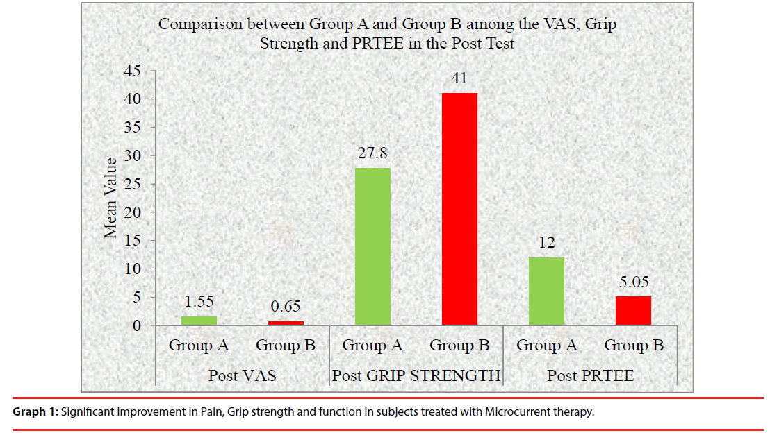
Graph 1 shows post test values of Visual Analogue Scale, grip strength and PRTEE Questionnaire between group A subjects treated with ULTRASOUND therapy and group B subjects treated with Microcurrent therapy. This graph shows there was a significant improvement in pain, grip strength and function among the subjects treated with MICROCURRENT than the other group of subjects treated with Ultrasound. Thus Microcurrent therapy is proved to be more effective than Ultrasound in subjects with chronic lateral epicondylitis.
Discussion
This study compares the effectiveness of Ultrasound therapy and Microcurrent therapy in subjects with chronic lateral epicondylitis. The study was done on 20 subjects divided into two groups Group A and Group B, 10 in each. Subjects in group A was treated with Ultrasound therapy and Subjects in group B was treated with Microcurrent therapy for 1 week each.
The statistical analysis performed between group A and group B showed the following outcomes. Group B treated with Microcurrent therapy for 1week showed a significant reduction in Pain, and increased in hand grip strength in subjects with chronic lateral epicondylitis with the statistical value of (P<0.05), then compared with group A subjects treated with Ultrasound therapy.
Microcurrent therapy reduces the pain improvement scores with accompanying substantial reduction in serum levels of the inflammatory cytokines IL-1, IL-6, and TNF-X, and neuropeptide substance P. Beta- endorphin release and increases in serum cortisol. It also promote the healing of tissues and inhibit the growth of various pathogens.
According to Lambert et al 2002 electro membrane Microcurrent therapy reduces signs and symptoms of muscle damage. Hand grip strength increased from pretest to post test in subjects which shows the statistical value at P<0.05. Reduction in pain at power-grip and lifting a weight load with pronated forearm, improvement in grip-strength in chronic lateral epicondylitis patients. Microcurrent therapy represents a significant improvement in rapid pain control and acceleration of healing. It uses currents in micro ampere range, and below sensation threshold.
Group A treated with ultrasound therapy shows a significant reduction in pain, increased grip strength and functional activities. Ultrasonic Therapy is a method of stimulating the tissue beneath the skin’s surface using very high frequency sound waves, between 800,000 Hz and 2,000,000 Hz. Ultrasound is a therapeutic modality that has been used by physical therapists since the 1940s. Ultrasound is applied using a round-headed wand or probe that is put in direct contact with the patient’s skin. Ultrasound gel is used on all surfaces of the head in order to reduce friction and assist in the transmission of the ultrasonic waves. Therapeutic ultrasound is in the frequency range of about 0.8-3.0 MHZ.
The waves are generated by a piezoelectric effect caused by the vibration of crystals within the head of the wand/probe. The sound waves that pass through the skin cause a vibration of the local tissues. This vibration or cavitation can cause a deep heating locally though usually no sensation of heat will be felt by the patient. It has been shown to cause increases in tissue relaxation, local blood flow, and scar tissue breakdown. The effect of the increase in local blood flow can be used to help reduce local swelling and chronic inflammation. It is suggested that the application of Ultrasound to injured tissues will, amongst other things, speed the rate of healing & enhance the quality of the repair. These physiological aspects clearly explain the reason over the reduction of pain following ultrasound management for a period of one week.
The posttest mean value of Pain [VAS], grip strength and functional activities of group A treated with Ultrasound therapy was 1.55 and 27.80 and group B treated with Microcurrent therapy was 0.65 and 41.0 at the end of 1 week simultaneously. Hence the recovery is faster, pain free and effective in group B subjects treated with Microcurrent therapy. Thus these statistical findings could be attributed to the fact that Microcurrent therapy works more statistically over ultrasound therapy.
Conclusion
The application of 1 week treatment of Microcurrent therapy had increased the muscle function through reduction of muscle tension and strengthening of the weakened muscles of patients with lateral epicondylitis.
Pain intensity and Functional disability are reduced significantly. Hand grip strength also increased from pretest to posttest. This study demonstrated the effects of Microcurrent in improving muscle endurance as well as in reducing pain symptoms and improving functional performance in patients with lateral epicondylitis.
Acknowledgment
The author was grateful to Prof. V.P.R Sivakumar, Dean, SRM College of Physiotherapy, SRM University for his valuable support and suggestions for completion of study.
References
- Kane SF, James MH, Taylor JC. Womack Army Medical Center, Fort Bragg, North Carolina -Evaluation of Elbow Pain in Adults.
- Garg R, Adamson GJ, Dawson PA, et al. A prospective randomized study comparing a forearm strap brace versus a wrist splint for the treatment of lateral epicondylitis. J. Shoulder Elbow Surg 19(4),508-512 (2010).
- Van Hofwegen C, Baker CL III, Baker CL Jr. Epicondylitis in the athlete’s elbow. Clin. Sports Med 29(4),577-597 (2010).
- Shiri R, Viikari‐Juntura E, Varonen H, et al.Prevalence and determinants of lateral and medial epicondylitis: a population study.Am. J. Epidemiol164,1064-1065 (2006).
- Conway JE, Jobe FW, Glousman RE, et al. Medial instability of the elbow in throwing athletes: treatment by repair or reconstruction of the UCL. J. Bone Joint Surg 74(1),67-83 (1992).
- Wright A, Vicenzino B. Lateral epicondylgia, Therapeutic management. Phys. Ther. 2(1),39-48 (1997).
- Wong AR, Schumann B, Townsend R, et al.A Survey of therapeutic ultrasound use by physical therapists who are orthopaedic certified specialists.Phys. Ther87(8), 986-994(2007).
- Poltawski L, Watson T. Bioelectricity and microcurrent therapy for tissue healing – a narrative review. Phys. Ther. Rev 14(2), 104-114 (2009).
- Lathrop HP. Physiological effects of microcurrent on the body.
- Lambert MI, Marcus P, Burgess T, et al. Electromembrane Microcurrent therapy reduces signs and symptoms of muscle damage. Med. Sci. Sports. Exerc 34(4), 602-607 (2002).
- Prentice W. Therapeutic modalities for sports medicine and athletic training (6thedn). Ther. Modalities McGrawHill 1(7) (2008).
- Watson T. Electrotherapy evidence based therapy. (12thedn) Churchill Livingstone, Elsevier (2008).
- Demir H, Menku P, Kirnap M, et al. Comparison of the effects of laser, ultrasound, and combined laser + ultrasound treatments in experimental tendon healing. Lasers Surg. Med 35(1), 84-89 (2004).
- Ng GY, Ng CO, See EK. Comparison of therapeutic ultrasound and exercises for augmenting tendon healing in rats. Ultrasound Med. Biol 30: 1539- 1543 (2004).
